Review
. 2016 Apr 27:4:29.
doi: 10.1186/s40560-016-0138-3. eCollection 2016.

Traumatic brain injury: pathophysiology for neurocritical care

Affiliations
Review

Traumatic brain injury: pathophysiology for neurocritical care

Kosaku Kinoshita. J Intensive Care. .

Abstract

Severe cases of traumatic brain injury (TBI) require neurocritical care, the goal being to stabilize hemodynamics and systemic oxygenation to prevent secondary brain injury. It is reported that approximately 45 % of dysoxygenation episodes during critical care have both extracranial and intracranial causes, such as intracranial hypertension and brain edema. For this reason, neurocritical care is incomplete if it only focuses on prevention of increased intracranial pressure (ICP) or decreased cerebral perfusion pressure (CPP). Arterial hypotension is a major risk factor for secondary brain injury, but hypertension with a loss of autoregulation response or excess hyperventilation to reduce ICP can also result in a critical condition in the brain and is associated with a poor outcome after TBI. Moreover, brain injury itself stimulates systemic inflammation, leading to increased permeability of the blood-brain barrier, exacerbated by secondary brain injury and resulting in increased ICP. Indeed, systemic inflammatory response syndrome after TBI reflects the extent of tissue damage at onset and predicts further tissue disruption, producing a worsening clinical condition and ultimately a poor outcome. Elevation of blood catecholamine levels after severe brain damage has been reported to contribute to the regulation of the cytokine network, but this phenomenon is a systemic protective response against systemic insults. Catecholamines are directly involved in the regulation of cytokines, and elevated levels appear to influence the immune system during stress. Medical complications are the leading cause of late morbidity and mortality in many types of brain damage. Neurocritical care after severe TBI has therefore been refined to focus not only on secondary brain injury but also on systemic organ damage after excitation of sympathetic nerves following a stress reaction.

Keywords: Catecholamine; Hyperglycemia; Neurocritical care; Pathophysiology; Traumatic brain injury.


Figures

Fig. 1
Fig. 1
Brain autoregulation (pressure regulation) curve. Cerebral blood flow (CBF) is constant when mean arterial blood pressure (MAP) is kept between 60 and 160 mmHg. As the cerebral vasculature changes to adjust to MAP, vasoconstriction or vasodilatation changes. In patients who had hypertension or severe traumatic brain injury (TBI), the autoregulation curve shifts to the right. Due to the rightward shift (arrow), a MAP-dependent CBF reduction (brain ischemia) or increase (hyperemia) occurs even for a small change in blood pressure. Note that the plateau range of CBF is presumably altered after TBI occurs. No clear data are available, however, on how this presumed alteration takes place
Fig. 2
Fig. 2
Vasodilation and vasoconstriction cascade in the cerebral vasculature. This cascade model was first described by Rosner in the 1990s (see references 22, 23). A cascade of this type is often trigged by changes in CPP. Any step in the cascade, however, can be triggered as the starting point. There are many triggering factors such as dehydration, vascular volume, systemic metabolism, CMRO2, blood viscosity, systemic oxygen delivery, PaCO2, or certain pharmacologic agents. SAP systemic arterial pressure, CPP cerebral perfusion pressure, ICP intracranial pressure, CBV cerebral blood volume, CMRO 2 cerebral metabolic rate for oxygen
Fig. 3
Fig. 3
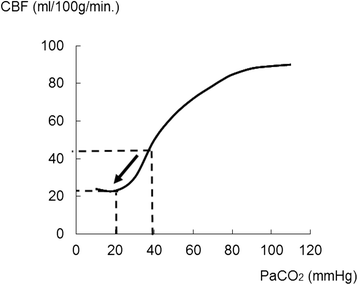
Changes in CBF related to PaCO2 level variation. In the case of respiratory acidosis, the effect of PaCO2 on the cerebral vasculature can augment cerebral blood flow (CBF). Conversely, CBF would be reduced by vasoconstriction after a drop in PaCO2. When PaCO2 values fall below 20 mmHg from about 40 mmHg, CBF also drops to half of the basic value (arrow)
Fig. 4
Fig. 4
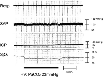
Brain ischemia after hyperventilation. A female in her 40s with traumatic brain injury was transferred to the hospital by ambulance. Brain CT scan revealed acute subdural hematoma. Surgical interventions were performed, and the patient’s ICP and SjO2 were monitored. The SjO2 value drops after hyperventilation. This phenomenon can be explained by the vasoconstriction effect from reduced PaCO2. Cerebral perfusion pressure changes might not have any remarkable effect because SAP and ICP values have been constant. Clinically, physicians would not be able to detect brain ischemia only from vital signs in this case without monitoring for brain oxygenation, such as SjO2 monitoring. The ICP will stay constant even if there are changes in the intracranial volume (e.g., the change in the volume of the vascular bed during the space compensatory phase). While the ICP will spread to the CSF space or any similar space until the compensatory effect is lost, no remarkable changes in the ICP are seen during the space compensatory phase. As a consequence, hyperventilation therapy for ICP control will not be effective in this phase. It may even cause harm via the decrease in CBF induced by excess vasoconstriction. Resp. respiration, SAP systemic arterial pressure, ICP intracranial pressure, SjO 2 jugular bulb oxygen saturation, HV hyperventilation. Data were obtained from brain injury patient monitored at our hospital in the 1990s
Fig. 5
Fig. 5
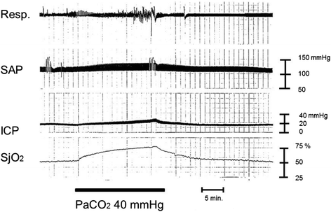
Effect on cerebral blood flow caused by augmentation of PaCO2. A male in his 30s suffered a traffic accident. Initial CT scan demonstrated acute subdural hematoma. Increased PaCO2 could stimulate the vasodilation cascade in the brain. As a result of an increase in PaCO2, the brain vasculature goes through vasodilation, with a subsequent increase in cerebral blood flow (and cerebral blood volume), leading to increased ICP. Physicians would be able to detect this from increased SjO2 in the clinical setting. Resp. respiration, SAP systemic arterial pressure, ICP intracranial pressure, SjO 2 jugular bulb oxygen saturation, CPP cerebral perfusion pressure. Data were obtained from brain injury patient monitored at our hospital in the 1990s
Fig. 6
Fig. 6
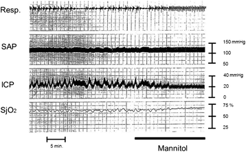
Effect of mannitol administration on patient with intracranial hypertension. A male in his 60s suffered traumatic brain injury. Brain CT scan demonstrated cerebral contusion. Mannitol administration is a potentially effective volume replacement method in the early phase and can stimulate the vasoconstriction cascade. SjO2 values gradually increase after mannitol administration. This phenomenon is likely caused by the volume expansion effect of mannitol, which could stimulate the vasoconstriction cascade leading to decreased CBV. Mannitol will then work as a hyperosmotic diuretic agent at the late phase resulting in decreased ICP and increased CPP. Resp. respiration, SAP systemic arterial pressure, ICP intracranial pressure, SjO 2 jugular bulb oxygen saturation, CBV cerebral blood volume, CPP cerebral perfusion pressure, Mannitol mannitol administration. Data were obtained from brain injury patient monitored at our hospital in the 1990s

References

    1. Gentile NT, McIntosh TK. Antagonists of excitatory amino acids and endogenous opioid peptides in the treatment of experimental central nervous system injury. Ann Emerg Med. 1993;22:1028–34. doi: 10.1016/S0196-0644(05)82746-5. - DOI - PubMed
    1. Faden AI, Demediuk P, Panter SS, Vink R. The role of excitatory amino acids and NMDA receptors in traumatic brain injury. Science. 1989;19(244):798–800. doi: 10.1126/science.2567056. - DOI - PubMed
    1. Xiong Y, Gu Q, Peterson PL, Muizelaar JP, Lee CP. Mitochondrial dysfunction and calcium perturbation induced by traumatic brain injury. J Neurotrauma. 1997;14:23–34. doi: 10.1089/neu.1997.14.23. - DOI - PubMed
    1. Yakovlev AG, Knoblach SM, Fan L, Fox GB, Goodnight R, Faden AI. Activation of CPP32-like caspases contributes to neuronal apoptosis and neurological dysfunction after traumatic brain injury. J Neurosci. 1997;17:7415–24. - PMC - PubMed
    1. Hall ED, Detloff MR, Johnson K, Kupina NC. Peroxynitrite-mediated protein nitration and lipid peroxidation in a mouse model of traumatic brain injury. J Neurotrauma. 2004;21:9–20. doi: 10.1089/089771504772695904. - DOI - PubMed

LinkOut - more resources