

Mechanisms of Perineural Invasion
- PMID: 27123385
- PMCID: PMC4846411
- DOI: 10.1055/s-0036-1571835
Mechanisms of Perineural Invasion
Abstract
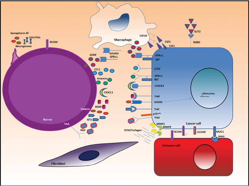
Perineural invasion (PNI) is the neoplastic invasion of nerves. PNI is widely recognized as an important adverse pathological feature of many malignancies, including pancreatic, prostate, and head and neck cancers and is associated with a poor prognosis. Despite widespread acknowledgment of the clinical significance of PNI, the mechanisms underlying its pathogenesis remain largely unknown. Recent theories of PNI pathogenesis have placed a significant emphasis on the active role of the nerve microenvironment, with PNI resulting from well-orchestrated reciprocal interactions between cancer and host. Elucidating the mechanisms involved in PNI may translate into targeted therapies for this ominous process.
Keywords: chemokine; head and neck cancer; neurotrophic factors; perineural invasion.
Figures

References
-
- Batsakis J G. Nerves and neurotropic carcinomas. Ann Otol Rhinol Laryngol. 1985;94(4 Pt 1):426–427. - PubMed
-
- Soo K C, Carter R L, O'Brien C J, Barr L, Bliss J M, Shaw H J. Prognostic implications of perineural spread in squamous carcinomas of the head and neck. Laryngoscope. 1986;96(10):1145–1148. - PubMed
-
- Fagan J J, Collins B, Barnes L, D'Amico F, Myers E N, Johnson J T. Perineural invasion in squamous cell carcinoma of the head and neck. Arch Otolaryngol Head Neck Surg. 1998;124(6):637–640. - PubMed
-
- Takahashi T, Ishikura H, Motohara T, Okushiba S, Dohke M, Katoh H. Perineural invasion by ductal adenocarcinoma of the pancreas. J Surg Oncol. 1997;65(3):164–170. - PubMed
-
- Cruveilheir J. Paris, France: JB Bailliere; 1835. Maladies Des Nerfs Anatomie Pathlogique Du Corps Humain.
Publication types
Grants and funding
LinkOut - more resources
Full Text Sources
Other Literature Sources
