Comparative Study
. 2016:2016:9128535.
doi: 10.1155/2016/9128535. Epub 2016 Mar 30.

Comparison of Calcium and Barium Microcapsules as Scaffolds in the Development of Artificial Dermal Papillae

Affiliations
Comparative Study

Comparison of Calcium and Barium Microcapsules as Scaffolds in the Development of Artificial Dermal Papillae

Yang Liu et al. Biomed Res Int. 2016.

Abstract

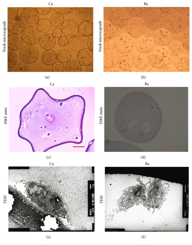
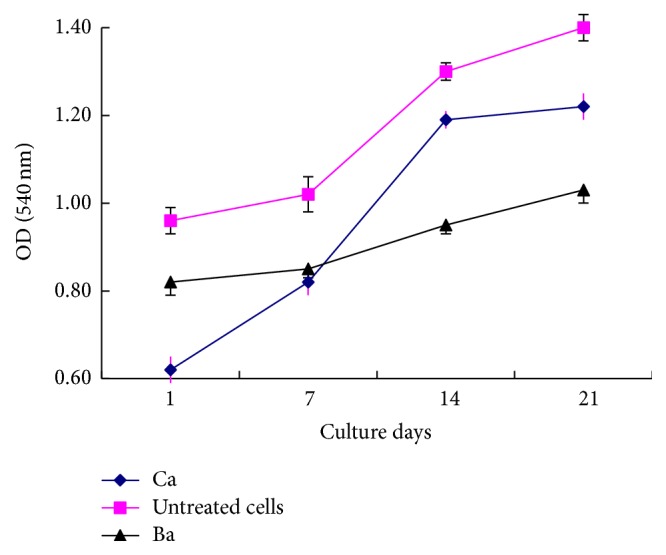
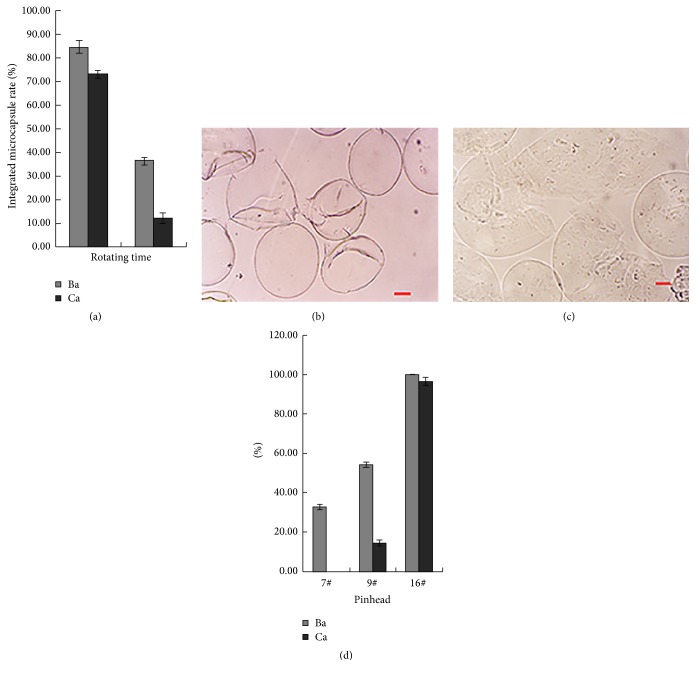
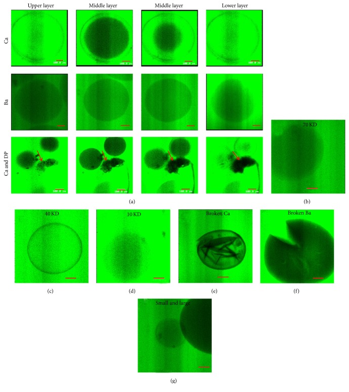
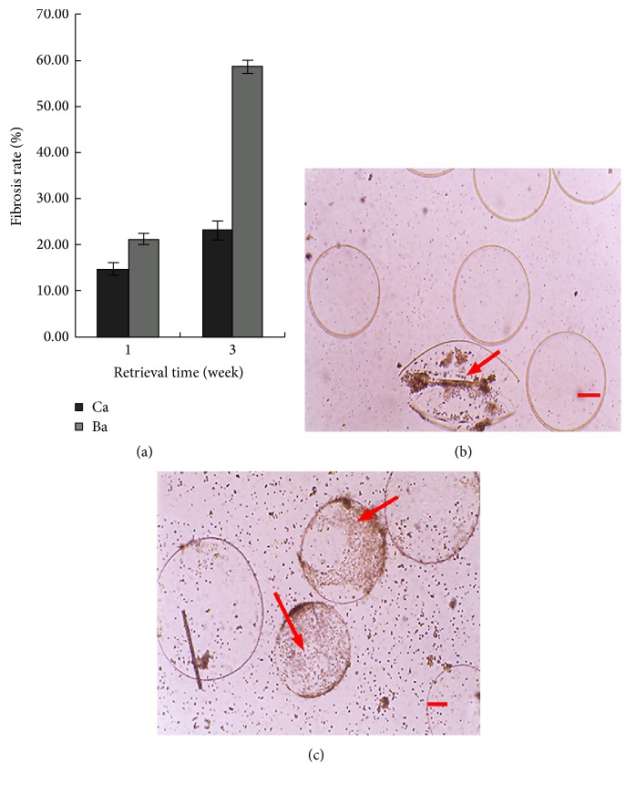
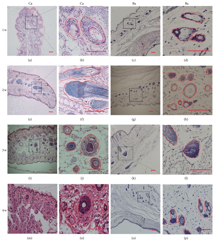
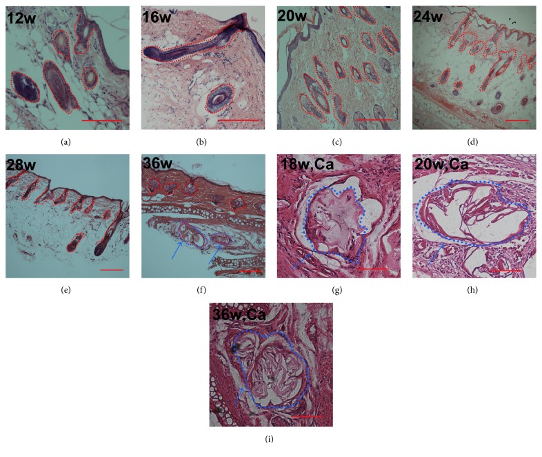
This study aimed to develop and evaluate barium and calcium microcapsules as candidates for scaffolding in artificial dermal papilla. Dermal papilla cells (DPCs) were isolated and cultured by one-step collagenase treatment. The DPC-Ba and DPC-Ca microcapsules were prepared by using a specially designed, high-voltage, electric-field droplet generator. Selected microcapsules were assessed for long-term inductive properties with xenotransplantation into Sprague-Dawley rat ears. Both barium and calcium microcapsules maintained xenogenic dermal papilla cells in an immunoisolated environment and induced the formation of hair follicle structures. Calcium microcapsules showed better biocompatibility, permeability, and cell viability in comparison with barium microcapsules. Before 18 weeks, calcium microcapsules gathered together, with no substantial immune response. After 32 weeks, some microcapsules were near inflammatory cells and wrapped with fiber. A few large hair follicles were found. Control samples showed no marked changes at the implantation site. Barium microcapsules were superior to calcium microcapsules in structural and mechanical stability. The cells encapsulated in hydrogel barium microcapsules exhibited higher short-term viability. This study established a model to culture DPCs in 3D culture conditions. Barium microcapsules may be useful in short-term transplantation study. Calcium microcapsules may provide an effective scaffold for the development of artificial dermal papilla.


Figures

Figure 1
Figure 1
Ca and Ba DPC microcapsule structure. Microcapsule structures were accessed with light microcopy (a, b). The packaged DP cells and extracellular structures were further studied with H&E staining (c, d). Transmission electron microscopy was utilized to observed detail structures of microcapsule membrane and inside DP cells (red circles) (e, f). Ca (a) and Ba (b) microcapsules appeared round, smooth, and transparent under inverted microscopy. H&E staining showed extracellular matrix secreted around cells (red arrow) (c). Ba microcapsules showed scattered cells at all times, with no extracellular matrix (d). TEM confirmed that DPCs adhered to the Ca microcapsule membrane in 24 to 72 hours, and the cells retained their fine structure (e). The membrane of the Ba microcapsules was smooth, with solid and homogeneous core and cells inside showed many microvilli (red arrows) (f). (Bar = 100 μm (a, b, c, and d), Bar = 1 μm (e, f).)
Figure 2
Figure 2
Viability and growth of embedded cells. Populations of alive cell in microencapsulated cells and untreated cells were determined by MTT assay. Data are represented as mean ± SD from 3 experiments.
Figure 3
Figure 3
Structural stability microcapsules. Thousands each of DPC-Ba and DPC-Ca microcapsules were assessed by determining change in structural integrity under an inverted microscope at 10 and 60 min after being stirred in flask (a). Cells inside the fragmented Ca microcapsules escaped (b). Cells were retained inside the membrane of Ba microcapsules, even after fragmentation (c). 1 mL of Ba and Ca microcapsules of 400 μm diameter were injected through 7#, 9#, and 16# pinheads, and integrated microcapsules were counted under a phase-contrast microscope (d). (Bar = 100 μm.)
Figure 4
Figure 4
Permeability of artificial and natural membranes. (a) The diffusion of fluorescence in Ca and Ba microcapsules and fresh DP (red arrow) was monitored by scanning from the center every 5 μm ×4 times by confocal microscopy after being permeabilized for 60 min. (b, c, d) The Ca microcapsules permeabilized for 30 minutes with different molecular weight of FITC-dextran. The medium and FITC-dextran entered the broken Ca capsules immediately (e) but not into Ba microcapsules even after fragmentation (f). (g) The fluorescence was stronger for 200 μm microcapsules compared with 600 μm diameter Ca microcapsules. (Bar = 100 μm.)
Figure 5
Figure 5
Biocompatibility of microcapsules. The same number of empty DPC-Ba and DPC-Ca microcapsules was introduced into the peritoneal cavity of mice and retrieved after 1 and 3 weeks for measurement of rate of fibrosis (a). One week after transplantation into the mouse peritoneal cavity, the retrieved Ca (b) and Ba (c) microcapsules showed increased fibrosis (2.3% and 5.9% (P < 0.01), resp.), with surrounding inflammatory cells. (Bar = 100 μm.)
Figure 6
Figure 6
Short-term study of DPC-Ca and DPC-Ba microcapsules transplanted into rat ears. Large hair follicles formed after Ca microcapsule implantation at weeks 1–4 (a, b, e, f, i, j, m, and n). At 2 weeks, large DP formed near the transplanted site without the surrounding DPC microcapsule (e, f). No abnormal hair follicle structures were found in Ba microcapsules at 1 to 2 weeks after transplantation (c, d, g, and h). At 3 to 4 weeks, Ba (k, l, o, and p) microcapsules produced large hair follicles but fewer compared with Ca (i, j, m, and n) microcapsules. (Bar = 100 μm.)
Figure 7
Figure 7
Long-term study of DPC-Ca microcapsules transplanted into rat ears and the histology of Ca microcapsules at different weeks. From 12 to 36 weeks, the number and size of large hair follicles decreased (a, b, c, d, e, and f). At 18 weeks, Ca microcapsules were found at the transplanted site, with no surrounding inflammatory cells (g). At 20 weeks, some inflammatory cells were around the microcapsules (h). At 36 weeks the number of inflammatory cells was reduced, with fibers around the microcapsules (i). (Bar = 100 μm.) Red circle: microcapsules and formed HFs. Blue circle and arrow: microcapsules surrounded associated with inflammatory cells.

Similar articles

References

    1. Birch M. P., Messenger A. G. Genetic factors predispose to balding and non-balding in men. European Journal of Dermatology. 2001;11(4):309–314. - PubMed
    1. Stough D., Stenn K., Haber R., et al. Psychological effect, pathophysiology, and management of androgenetic alopecia in men. Mayo Clinic Proceedings. 2005;80(10):1316–1322. doi: 10.4065/80.10.1316. - DOI - PubMed
    1. Horne K. A., Jahoda C. A., Oliver R. F. Whisker growth induced by implantation of cultured vibrissa dermal papilla cells in the adult rat. Journal of Embryology and Experimental Morphology. 1986;97:111–124. - PubMed
    1. Inamatsu M., Matsuzaki T., Iwanari H., Yoshizato K. Establishment of rat dermal papilla cell lines that sustain the potency to induce hair follicles from afollicular skin. The Journal of Investigative Dermatology. 1998;111(5):767–775. doi: 10.1046/j.1523-1747.1998.00382.x. - DOI - PubMed
    1. Jahoda C. A. B., Horne K. A., Oliver R. F. Induction of hair growth by implantation of cultured dermal papilla cells. Nature. 1984;311(5986):560–562. doi: 10.1038/311560a0. - DOI - PubMed

Publication types

LinkOut - more resources