. 2016 Apr 28;11(4):e0153757.
doi: 10.1371/journal.pone.0153757. eCollection 2016.

Genetic Analysis of 'PAX6-Negative' Individuals with Aniridia or Gillespie Syndrome

Affiliations

Genetic Analysis of 'PAX6-Negative' Individuals with Aniridia or Gillespie Syndrome

Morad Ansari et al. PLoS One. .

Abstract

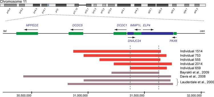
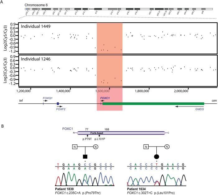
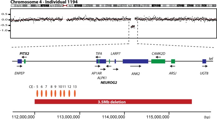
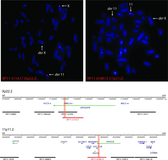
We report molecular genetic analysis of 42 affected individuals referred with a diagnosis of aniridia who previously screened as negative for intragenic PAX6 mutations. Of these 42, the diagnoses were 31 individuals with aniridia and 11 individuals referred with a diagnosis of Gillespie syndrome (iris hypoplasia, ataxia and mild to moderate developmental delay). Array-based comparative genomic hybridization identified six whole gene deletions: four encompassing PAX6 and two encompassing FOXC1. Six deletions with plausible cis-regulatory effects were identified: five that were 3' (telomeric) to PAX6 and one within a gene desert 5' (telomeric) to PITX2. Sequence analysis of the FOXC1 and PITX2 coding regions identified two plausibly pathogenic de novo FOXC1 missense mutations (p.Pro79Thr and p.Leu101Pro). No intragenic mutations were detected in PITX2. FISH mapping in an individual with Gillespie-like syndrome with an apparently balanced X;11 reciprocal translocation revealed disruption of a gene at each breakpoint: ARHGAP6 on the X chromosome and PHF21A on chromosome 11. In the other individuals with Gillespie syndrome no mutations were identified in either of these genes, or in HCCS which lies close to the Xp breakpoint. Disruption of PHF21A has previously been implicated in the causation of intellectual disability (but not aniridia). Plausibly causative mutations were identified in 15 out of 42 individuals (12/32 aniridia; 3/11 Gillespie syndrome). Fourteen of these mutations presented in the known aniridia genes; PAX6, FOXC1 and PITX2. The large number of individuals in the cohort with no mutation identified suggests greater locus heterogeneity may exist in both isolated and syndromic aniridia than was previously appreciated.


Conflict of interest statement

Competing Interests: The authors have declared that no competing interests exist.

Figures

Fig 1
Fig 1. Identification of PAX6 whole-gene deletions.
Genome-wide array CGH analysis identified a 650 kb deletion in individual 2193 (chr11:31,199,000–31,849,000), a 520 kb deletion in individual 377 (chr11:31,394,000–31,914,000), a 154 kb deletion in individual 1510 (chr11:31,779,000–31,933,000) and a 96 kb deletion in individual 1977 (chr11:31,698,271–31,794,414), all involving PAX6. Red bars show the position of the deletions. Genes transcribed on the forward strand are in blue and those transcribed on the reverse strand are in green, also indicated by arrows. Genomic coordinates are based on the Human Genome Assembly hg18.
Fig 2
Fig 2. Identification of regulatory deletions telomeric to PAX6.
Regulatory deletions telomeric to PAX6 were identified in individual 1514 (chr11:30,874,642–31,654,833), individual 753 (chr11:30,967,000–31,704,000), individual 555 (chr11:31,108,579–31,649–842), individual 2014 (chr11:31,234,395–31,751,815) and individual 659 (chr11:31,379,000–31,708,000). The schematic diagram shows how the ‘critical region’ (delimited by grey dotted lines) required for PAX6 transcriptional activation was delineated by combining our data with published deletions with known coordinates [55,67,68]. PAX6 regulatory deletions from the present study are shown by red blocks. Genes transcribed on the forward strand are in blue and those transcribed on the reverse strand are in green, also indicated by arrows. Genomic coordinates are based on the Human Genome Assembly hg18.
Fig 3
Fig 3. Mutation analysis of the FOXC1 locus.
(A) Genome-wide array CGH identified two deletions encompassing the FOXC1 gene in individuals 1449 (chr6:1,543,591–1,675,085) and 1246 (chr6:1,543,591–1,675,085). (B) Direct sequencing of the FOXC1 coding region identified a heterozygous substitution in individual 1839 (c.235C>A, p.(Pro79Thr)) and another in individual 1634 (c.302T>C, p.(Leu101Pro)). FOXC1 mutation screening in unaffected parents of both patients showed that the mutations had occurred de novo. The locations of both mutations within the fork-head domain of the FOXC1 protein are indicated by vertical arrows. Genes transcribed on the forward strand are in blue and those transcribed on the reverse strand are in green, also indicated by arrows. Genomic coordinates are based on the Human Genome Assembly hg18. The genomic sequence identifier for FOXC1 is NG_009368.
Fig 4
Fig 4. Identification of a potential PITX2 regulatory deletion.
Genome-wide array CGH identified a deletion of approximately 3.5 Mb in individual 1194 (chr4:111,994,000–115,504,000) (red bar). The deletion is located telomeric to the PITX2 gene on chromosome 4. The positions of conserved elements (CE) in the deleted region, as identified by Volkmann et al., 2011 [47] are marked by orange ellipses. Genes transcribed on the forward strand are in blue and those transcribed on the reverse strand are in green, also indicated by arrows. Genomic coordinates are shown on the x-axis and are based on the Human Genome Assembly hg18.
Fig 5
Fig 5. Fluorescence in situ hybridization (FISH) was used to map the translocation breakpoints on chromosomes 11 and X in individual 1371.
The breakpoint-spanning BAC clones RP11-311A17 (Xp22.2; left panel) and RP11-618K13 (11p11.2; right panel) show signals on both the derivative 11 and derivative X. The schematic diagram demonstrates the position of the BAC clones and the genes involved, to scale. Breakpoint-spanning BACs are coloured in red, with the approximate position of the breakpoints shown by orange bars, as determined by long-range PCR. Genes transcribed on the forward strand are in blue and those transcribed on the reverse strand are in green. Genomic coordinates are shown on the x-axis and are based on the Human Genome Assembly hg18.

References

    1. Boonstra N, Limburg H, Tijmes N, van Genderen M, Schuil J, van Nispen R (2012) Changes in causes of low vision between 1988 and 2009 in a Dutch population of children. Acta Ophthalmol 90: 277–286. 10.1111/j.1755-3768.2011.02205.x - DOI - PubMed
    1. Gronskov K, Rosenberg T, Sand A, Brondum-Nielsen K (1999) Mutational analysis of PAX6: 16 novel mutations including 5 missense mutations with a mild aniridia phenotype. Eur J Hum Genet 7: 274–286. - PubMed
    1. Dubey SK, Mahalaxmi N, Vijayalakshmi P, Sundaresan P (2015) Mutational analysis and genotype-phenotype correlations in southern Indian patients with sporadic and familial aniridia. Mol Vis 21: 88–97. - PMC - PubMed
    1. Hever AM, Williamson KA, van Heyningen V (2006) Developmental malformations of the eye: the role of PAX6, SOX2 and OTX2. Clin Genet 69: 459–470. - PubMed
    1. Hingorani M, Hanson I, van Heyningen V (2012) Aniridia. Eur J Hum Genet 20: 1011–1017. 10.1038/ejhg.2012.100 - DOI - PMC - PubMed

Publication types

MeSH terms

Supplementary concepts