

Embryologic and Fetal Development of the Human Eyelid
- PMID: 27124372
- PMCID: PMC5102278
- DOI: 10.1097/IOP.0000000000000702
Embryologic and Fetal Development of the Human Eyelid
Abstract
Purpose: To review the recent data about eyelid morphogenesis, and outline a timeline for eyelid development from the very early stages during embryonic life till final maturation of the eyelid late in fetal life.
Methods: The authors extensively review major studies detailing human embryologic and fetal eyelid morphogenesis. These studies span almost a century and include some more recent cadaver studies. Numerous studies in the murine model have helped to better understand the molecular signals that govern eyelid embryogenesis. The authors summarize the current findings in molecular biology, and highlight the most significant studies in mice regarding the multiple and interacting signaling pathways involved in regulating normal eyelid morphogenesis.
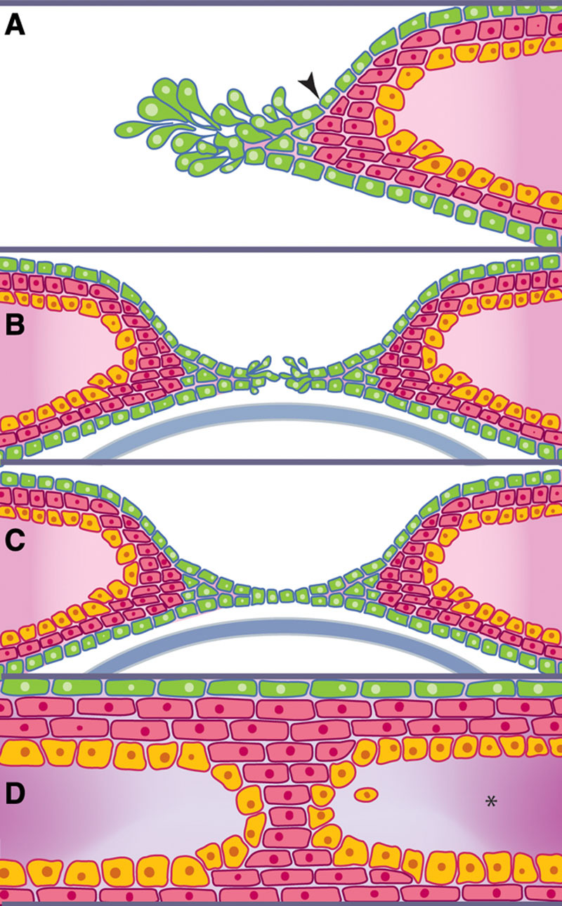
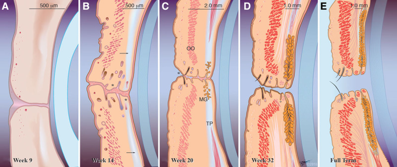
Results: Eyelid morphogenesis involves a succession of subtle yet strictly regulated morphogenetic episodes of tissue folding, proliferation, contraction, and even migration, which may occur simultaneously or in succession.
Conclusions: Understanding the extraordinary process of building eyelid tissue in embryonic life, and deciphering its underlying signaling machinery has far reaching clinical implications beyond understanding the developmental abnormalities involving the eyelids, and may pave the way for achieving scar-reducing therapies in adult mammalian wounds, or control the spread of malignancies.
Conflict of interest statement
The authors have no financial or conflicts of interest to disclose.
Figures
References
-
- Gasser R. The virtual human embryo. Available at: http://virtualhumanembryo.lsuhsc.edu/. Accessed October 24, 2015.
-
- Salomon LJ, Alfirevic Z, Bilardo CM, et al. ISUOG practice guidelines: performance of first-trimester fetal ultrasound scan. Ultrasound Obstet Gynecol. 2013;41:102–13. - PubMed
-
- Schoenwolf G, Bleyl S, Brauer P, Francis-West P. Origin of epidermis and dermis of skin. In: Schoenwolf G, Bleyl S, Brauer P, Francis-West P, editors. In: Larsen’s Human Embryology. 4th ed. Philadelphia, PA: Churchill Livingstone Elsevier; 2009. Available at: https://www.inkling.com/read/larsens-human-embryology-schoenwolf-bleyl-b.... Accessed November 5, 2013.
-
- Smyth I, Scambler P. The genetics of Fraser syndrome and the blebs mouse mutants. Hum Mol Genet. 2005;14 Spec No. 2:R269–74. - PubMed
Publication types
MeSH terms
LinkOut - more resources
Full Text Sources
Other Literature Sources
Medical
