. 2016 Apr 28;7(4):e2210.
doi: 10.1038/cddis.2016.84.

MicroRNA-682-mediated downregulation of PTEN in intestinal epithelial cells ameliorates intestinal ischemia-reperfusion injury

Affiliations

MicroRNA-682-mediated downregulation of PTEN in intestinal epithelial cells ameliorates intestinal ischemia-reperfusion injury

Z Liu et al. Cell Death Dis. .

Abstract

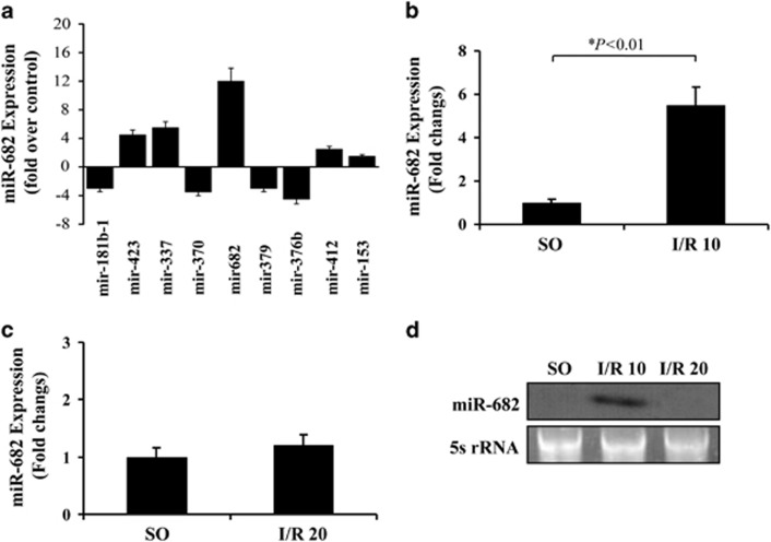
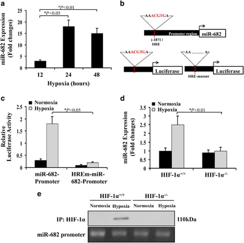
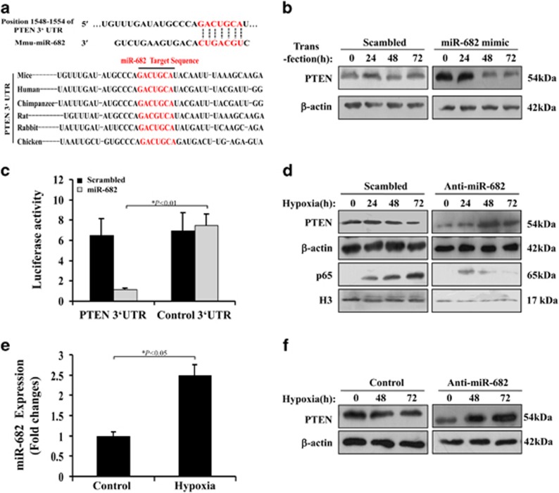
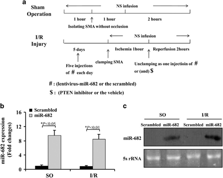
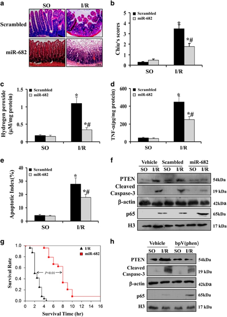
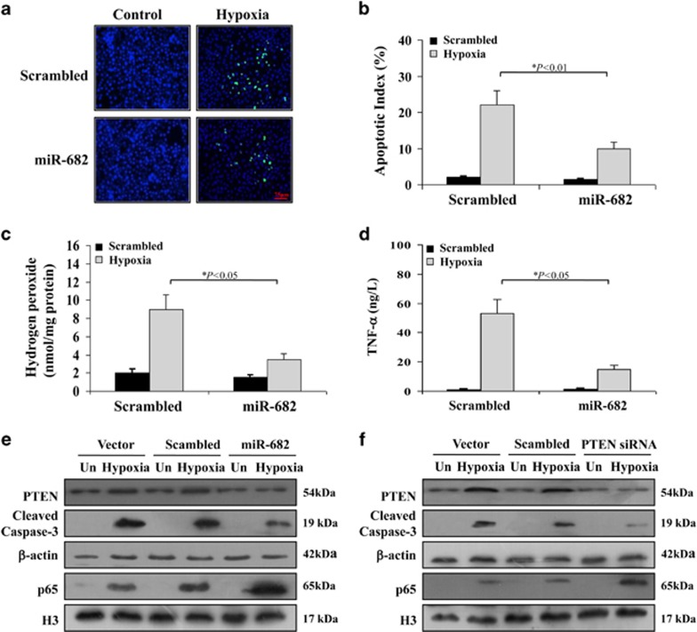
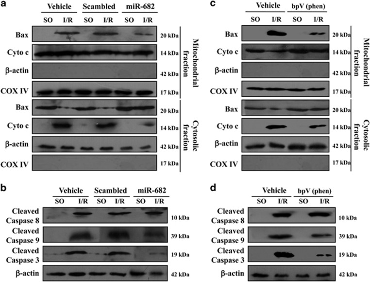
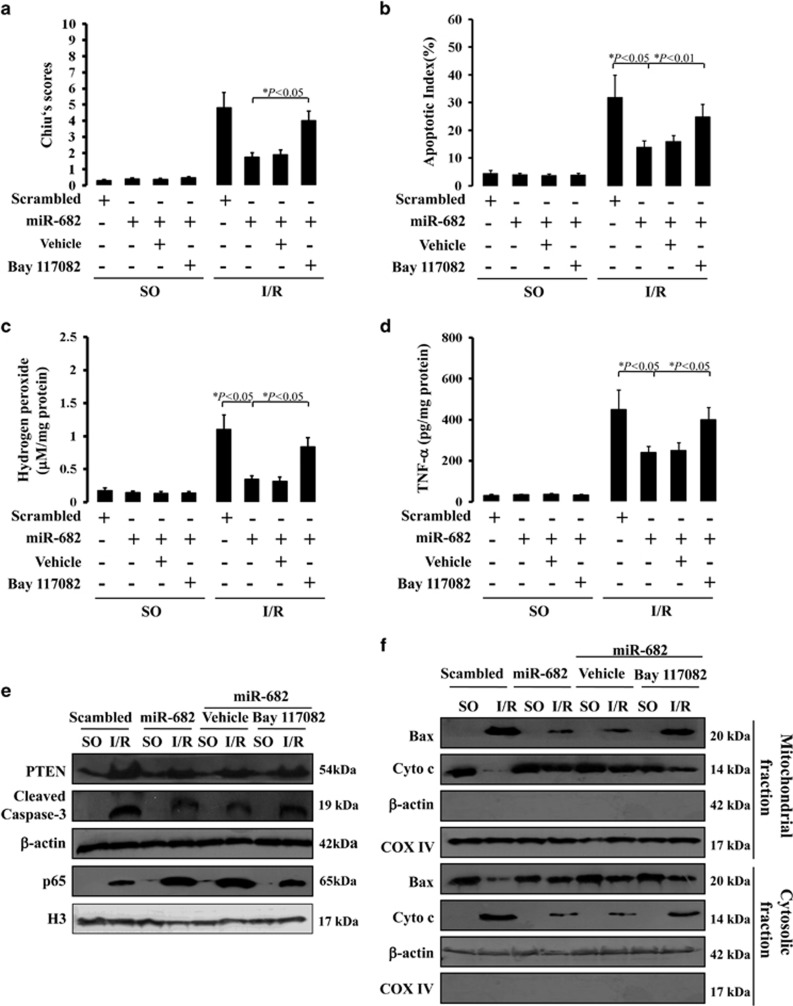
Intestinal ischemia-reperfusion (I/R) injury causes inflammation and tissue damage and contributes to high morbidity and mortality, but the underlying mechanism remains elusive and effective therapies are still lacking. We report here a critical role of the microRNA 682 (miR-682) as a key regulator and therapeutic target in intestinal I/R injury. MiR-682 was markedly induced in intestinal epithelial cells (IECs) during intestinal ischemia in mice and in the human colonic epithelial cells during hypoxia, but was undetected rapidly after intestinal reperfusion in IEC of mice. MiR-682 induction during hypoxia was modulated by hypoxia-inducible factor-1α (HIF-1α). On lentivirus-mediated miR-682 overexpression in vivo during intestinal reperfusion or miR-682 mimic transfection in vitro during hypoxia, miR-682 decreased the expression of phosphatase and tensin homolog (PTEN) and subsequently activated nuclear translocation of nuclear factor kappa B (NF-κB) p65. Consequently, NF-κB activation by miR-682-mediated PTEN downregulation prevented reactive oxygen species (ROS) induction, inflammatory reaction, mitochondrial-mediated apoptosis and IEC apoptosis. The effect of miR-682-mediated PTEN/NF-κB pathway on IECs resulted in protection against intestinal I/R injury in mice. However, NF-κB chemical inhibitor reversed miR-682-mediated decreased PTEN expression, ROS induction, inflammation and IEC apoptosis. Collectively, these results identify a novel miR-682/PTEN/NF-κBp65 signaling pathway in IEC injury induced by I/R that could be targeted for therapy.


Figures

Figure 1
Figure 1
Upregulation of miR-682 in IEC of mice during intestinal ischemia. (a) Significant alteration in miRNA expression in IEC of mice during intestinal ischemia. Mice were subjected to 1 h of the superior mesenteric artery ischemia, while control mice had sham operation (SO). Values are means±S.D., n=10 in each group. Four independent experiments were performed. (b) Real-time PCR of miR-682. RNA from IEC isolated from sham-operated control mice or mice with ischemia following by 10:min of reperfusion (I/R 10). Values are means±S.D., n=6 in each group. Three independent experiments were performed. (c) Real-time PCR of miR-682. RNA from IEC isolated from sham-operated control mice or mice with ischemia following by 20 min of reperfusion (I/R 20). Values are means±S.D., n=6 in each group. Three independent experiments were performed. (d) Northern blot analysis of miR-682. Ten micrograms of the total RNA extracted from IEC isolated from sham-operated control mice or mice with ischemia following by 10 or 20 min of reperfusion were used for Northern blot analysis. 5s r-RNA was probed as loading control
Figure 2
Figure 2
HIF-1 mediates miR-682 induction during hypoxia. (a) Induction of miR-682 by hypoxia. CCC-HIE-2 cells were incubated under hypoxia (1% oxygen) for 12–48 h to extract RNA for real-time PCR analysis of miR-682. Fold changes relative to cells without hypoxia treatment. Three independent experiments were performed. (b) Upper: miR-682 promoter region harboring HRE- or HIF-binding site. Lower: promoter reporter vectors containing the miR-682 promoter with or without HRE upstream of luciferase gene. (c) Activation of miR-682 promoter by hypoxia. miR-682 promoter or its HIF-binding site-deletion mutant were subcloned upstream of the luciferase gene in the promoter reporter construct. CCC-HIE-2 cells were contransfected with one of these reporter constructs, along with the Renilla luciferase construct, in a ratio of 2:0.1 and were then subjected to 24 h of hypoxia to collect lysate to measure luciferase activities. Four independent experiments were performed. (d) Induction of miR-682 expression by hypoxia in HIF-1α+/+ MEFs, but not in HIF-1α−/−cells. The cells were subjected to 24 h of hypoxia to isolated RNA for real-time PCR analysis of miR-682. Four independent experiments were performed. (e) HIF-1 binding to miR-682 promoter during hypoxia. HIF-1α+/+ and HIF-1α−/− cells were incubated under hypoxia or normoxia for 24 h. Cell lysate was collected for ChIP analysis of HIF-1 binding to miR-682 promoter DNA. Three independent experiments were performed. MEF, mouse embryonic fibroblast
Figure 3
Figure 3
miR-682 targets PTEN during hypoxic condition. (a) Upper: putative miR-682 complementary sequence in the 3′UTR of murine PTEN mRNA. Lower: conserved miR-682 target sequence in the PTEN 3′UTR. (b) CCC-HIE-2 cells transfected with a control Scrambled or miR-682 mimic, and western blot analysis was carried out from the whole cell lysates collected at different time points. Four independent experiments were performed. (c) Luciferase reporter assay was conducted using constructs with the PTEN 3′UTR or an antisense control sequence. CCC-HIE-2 cells were cotransfected with these constructs along with the scrambled or miR-682 mimic. Four independent experiments were performed. (d) CCC-HIE-2 cells transfected with a control Scrambled or anti-miR-682 LNA were subjected to hypoxia, and whole cell lysates were collected at indicated time points. Four independent experiments were performed. (e) Quantitative PCR analysis of miR-682 gene expression in CCC-HIE-2 cells treated under hypoxia condition after 12 h. (f) IEC-6 cells transfected with a control scrambled LNA or anti-miR-682 LNA were subjected to hypoxia, and whole cell lysates were collected at indicated time points. Western blot analysis of these lysates. Four independent experiments were performed
Figure 4
Figure 4
miR-682 protects small intestine against ischemia–reperfusion injury through PTEN/NF-κB p65 pathway. (a) The protocol of experiments. Sham-operated (SO) group: involving isolation of the superior mesenteric artery (SMA) without occlusion; ischemia–reperfusion Injury (I/R) group and treatments: performed by 1 h occlusion of SMA followed by 2 h of reperfusion without intervention. (b) Quantitative PCR analysis of miR-682 gene expression in isolated IEC cells of mice treated with or without I/R injury with a scrambled control or lentivirus-miR-682 (miR-682). Ten independent experiments were performed. (c) Northern blot analysis of miR-682 expression of IEC of mice after six i.v. injections of lentivirus-miR-682 (miR-682) with or without I/R injury. Forty micrograms of the total RNA extracted from isolated IEC were used for Northern blot analysis. 5 s r-RNA was probed as loading control. Six independent experiments were performed
Figure 5
Figure 5
miR-682 protects small intestine against ischemia–reperfusion injury through PTEN/NF-κB p65 pathway. (a) H&E staining was performed using formalin-fixed tissue sections from sham-operated control (SO) mice or (I/R) mice with 60 min of ischemia following by 120 min of reperfusion with or without pretreatment with lentivirus-miR-682 (miR-682) or a scrambled control. Magnification: × 400. (b) Chiu′s scores were measured and compared by ANOVA with Tukey post test. *P<0.05 versus SO. #P<0.05 versus Scrambled. Values are means±S.D., n=6 in each group. Three independent experiments were performed. (c) Levels of hydrogen peroxide were measured in IEC isolated from SO mice and I/R mice with or without pretreatment of lentivirus-miR-682 (miR-682) or a scrambled control. *P<0.01 versus SO. #P<0.05 versus Scrambled. Values are means±S.D., n=6 in each group. Three independent experiments were performed. (d) ELISA analysis of TNF-α protein expression in the IEC isolated from SO mice and I/R mice with or without pretreatment of lentivirus-miR-682 (miR-682) or a scrambled control. *P<0.01 versus SO. #P<0.01 versus Scrambled. Values are means±S.D., n=6 in each group. Three independent experiments were performed. (e) Apoptotic index was measured by counting a minimum of 20 randomly selected villi and crypts in the sections following TUNEL staining. The index was obtained by dividing the TUNEL-positive cells by the total number of cells. *P<0.05 versus SO. #P<0.05 versus Scrambled. Values are means±S.D., n=6 in each group. Three independent experiments were performed. (f) Western blot analysis of the IEC isolated from SO mice and I/R mice with or without pretreatment of lentivirus-miR-682 (miR-682) or a scrambled control. β-actin and nuclear H3 were used as the controls for loading. (g) Survival curves of I/R mice with or without pretreatment of lentivirus-miR-682 (miR-682). Values are means±S.D., n=8 in each group. Three independent experiments were performed. (h) Western blot analysis of the IEC isolated from SO mice and I/R mice with or without pretreatment of PTEN inhibitor bpV(phen) or vehicle. β-actin and nuclear H3 were used as the controls for loading
Figure 6
Figure 6
miR-682 suppresses hypoxia-induced cell apoptosis via PTEN/NF-κB p65 pathway. (a) TUNEL fluorescent staining in CCC-HIE-2 cells subjected to hypoxia after 24 h, with or without pretreatment with miR-682 mimic or a scrambled control. Magnification: × 400. (b) The apoptotic index was calculated by counting a minimum of 20 randomly selected fields following TUNEL staining. The index was obtained by dividing the TUNEL-positive cells by the total number of cells. Values are means±S.D.. Three independent experiments were performed. (c) Levels of hydrogen peroxide were measured in CCC-HIE-2 cells under hypoxia conditions after 24 h with or without pretreatment of miR-682 mimic or a scrambled control. Three independent experiments were performed. (d) ELISA analysis of TNF-α protein expression in CCC-HIE-2 cells under hypoxia conditions after 24 h with or without pretreatment of miR-682 mimic or a scrambled control. Three independent experiments were performed. (e) Western blot analysis of CCC-HIE-2 cells subjected to hypoxia after 24 h with or without pretreatment of miR-682 mimic or a scrambled control. β-actin nuclear H3 was used as the control for loading. Three independent experiments were performed. (f) Western blot analysis of CCC-HIE-2 cells subjected to hypoxia after 24 h with or without pretreatment of PTEN siRNA or a scrambled control. β-actin nuclear H3 was used as the control for loading. Three independent experiments were performed
Figure 7
Figure 7
miR-682 represses mitochondrial-mediated apoptosis in IEC of mice in response to intestinal I/R injury (a) Mitochondrial and cytosolic fractions were analyzed for Bax and cytochrome c by western blotting. β-actin and COX IV were markers of cytosolic and mitochondrial fractions, respectively. Three independent experiments were performed. (b) Western blot analysis of caspase-8, -9 and -3 activity of IEC cells isolated from sham-operated control (SO) mice or (I/R) mice with or without pretreatment with lentivirus-miR-682 (miR-682) or a scrambled control. (c) Mitochondrial and cytosolic fractions were analyzed for Bax and cytochrome c by western blotting. β-actin and COX IV were markers of cytosolic and mitochondrial fractions, respectively. Three independent experiments were performed. (d) Western blot analysis of caspase-8, -9 and -3 activity of IEC from sham-operated control (SO) mice or (I/R) mice with or without pretreatment with PTEN inhibitor bpV(phen) or vehicle
Figure 8
Figure 8
NF-κB inhibitor compromises intestinal protection of miR-682 following intestinal I/R injury. (a) Chiu′s scores were measured and compared by ANOVA with Tukey post test. Values are means±S.D., n=6 in each group. Three independent experiments were performed. (b) Apoptotic index in the crypts measured by TUNEL staining. Values are means±S.D., n=6 in each group. Three independent experiments were performed. (c) Levels of hydrogen peroxide were measured in IEC isolated from SO mice and I/R mice with pretreatment of lentivirus-miR-682 (miR-682) or a scrambled control with or without treatment of Bay117082. Values are means±S.D., n=6 in each group. Three independent experiments were performed. (d) ELISA analysis of TNF-α protein expression in IEC isolated from SO mice and I/R mice with pretreatment of lentivirus-miR-682 (miR-682) or a scrambled control with or without treatment of Bay117082. Values are means±S.D., n=6 in each group. Three independent experiments were performed. (e) Western blot analysis of IEC isolated from SO mice and I/R mice with pretreatment of lentivirus-miR-682 (miR-682) or a scrambled control with or without treatment of Bay117082. β-actin and nuclear H3 were used as the controls for loading. Three independent experiments were performed. (f) Western blot analysis of mitochondrial and cytosolic fractions from IEC isolated from SO mice and I/R mice with pretreatment of lentivirus-miR-682 (miR-682) or a scrambled control with or without treatment of Bay117082. β-actin and COX IV were markers of cytosolic and mitochondrial fractions, respectively. Three independent experiments were performed

References

    1. Mendell JT, Olson EN. MicroRNAs in stress signaling and human disease. Cell 2012; 148: 1172–1187. - PMC - PubMed
    1. Bartel DP. MicroRNAs: target recognition and regulatory functions. Cell 2009; 136: 215–233. - PMC - PubMed
    1. Ambros V. The functions of animal microRNAs. Nature 431: 350–355. - PubMed
    1. Schickel R, Boyerinas B, Park SM, Peter ME. (2008) MicroRNAs: key players in the immune system, differentiation, tumorigenesis and cell death. Oncogene 2004; 27: 5959–5974. - PubMed
    1. Ma L, Teruya-Feldstein J, Weinberg RA. Tumour invasion and metastasis initiated by microRNA-10b in breast cancer. Nature 2007; 449: 682–688. - PubMed

Publication types

MeSH terms