. 2016 Apr 28;11(4):e0154461.
doi: 10.1371/journal.pone.0154461. eCollection 2016.

Influenza-Specific Antibody-Dependent Phagocytosis

Affiliations

Influenza-Specific Antibody-Dependent Phagocytosis

Fernanda Ana-Sosa-Batiz et al. PLoS One. .

Abstract

Background: Immunity to human influenza A virus (IAV) infection is only partially understood. Broadly non-neutralizing antibodies may assist in reducing disease but have not been well characterized.

Methods: We measured internalization of opsonized, influenza protein-coated fluorescent beads and live IAV into a monocytic cell line to study antibody-dependent phagocytosis (ADP) against multiple influenza hemagglutinin (HA) subtypes. We analyzed influenza HA-specific ADP in healthy human donors, in preparations of intravenous immunoglobulin (IVIG), and following IAV infection of humans and macaques.

Results: We found that both sera from healthy adults and IVIG preparations had broad ADP to multiple seasonal HA proteins and weak cross-reactive ADP to non-circulating HA proteins. The ADP in experimentally influenza-infected macaque plasma and naturally influenza-infected human sera mediated phagocytosis of both homologous and heterologous IAVs. Further, the IAV phagocytosed in an antibody-mediated manner had reduced infectivity in vitro.

Conclusion: We conclude that IAV infections in humans and macaques leads to the development of influenza-specific ADP that can clear IAV infection in vitro. Repeated exposure of humans to multiple IAV infections likely leads to the development of ADP that is cross-reactive to strains not previously encountered. Further analyses of the protective capacity of broadly reactive influenza-specific ADP is warranted.


Conflict of interest statement

Competing Interests: Dr. Steven Rockman is an employee of bioCSL Ltd, a subsidiary of CSL Ltd that manufactures IVIG. The remaining authors do not have a commercial or other association that might pose a conflict of interest. This does not alter the authors' adherence to PLOS ONE policies on sharing data and materials.

Figures

Fig 1
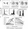
Fig 1. Representative influenza HA-specific ADP assay.
The ADP assay is performed as describe in (A). In brief, FITC fluorescent beads coated with hemagglutinin (HA) and fluorescent internalization probe (FIPCy5) are opsonized with 10μg/ml purified IgG from blood plasma or sera. HA-coated beads are incubated 16 hr with monocyte-like cell line, THP-1. Cy5 fluorescence from surface bound beads is quenched. Cells are fixed and analysed by flow cytometry. (B) Gating strategy of single cells. (C) Gating strategy for internalized beads+ (FITC+, Cy5+) and surface associate beads+ (FITC+. Cy5-) cells. Uptake of H3N2 A/Wyoming/03/2003 HA-coated beads in presence of influenza sero-negative human serum IgG or healthy human adult plasma IgG (top 2 panels). Uptake of H1N1 A/Puerto Rico/08/1939 HA-coated beads opsonized with IgG from macaque plasma either pre-influenza infection or post-influenza infection (bottom 2 panels). Data are representative of 14 human donors (Fig 2) and 6 macaques (Fig 5).
Fig 2
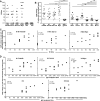
Fig 2. Comparison of HI titres and ADP activity in healthy adults.
(A) H1N1 A/California/04/2009 (H1N1 Pdm09), A/Solomon Islands/03/2006 (H1N1 Sol Isl), A/New Caledonia/20/1999 (H1N1 New Cal), H3N2 A/Wyoming/03/2003 (H3N2 Wyo), H5N1 A/Vietnam/1194/2004 (H5N1 Viet) γ-irradiated and H7N9 A/Anhui/01/2013 (H7N9 Anh) γ-irradiated hemagglutination inhibition (HI) titres were determined for 14 healthy donors. (B) ADP activity in serum IgG against seasonal H1N1 and H3N2 HAs (left panel) and rare H2N2 A/Japan/503/1957 (H2 Japan), H4H6 A/Swine/Ontario/01911-1/1999 (H4 Ontario), H5N1 A/Vietnam/1194/2004 (H5 Viet), H7N9 A/Shanghai/1/2013 (H7 Shan) and H7N7 A/Netherlands/219/2003 (H7 Neth) HAs (right panel) was evaluated. HIV-1 gp140 was included as negative control in each experiment. Line and error bars represent mean ± SEM. **** P < 0.0001, *** P < 0.001, ** P < 0.01, and ns P > 0.05 determined using Bonferroni’s multiple comparison test. Dotted line indicated mean + twice SD of ADP for HIV-1 gp140. (C) Correlation of H1 Pdm09, H1 Sol Isl and H3 Wyo HA-specific ADP activity and HI titre to the same virus determined using Pearson’s correlation. Data are representative of n = 3 independent experiments. (D) Correlation of H1 Pdm09, H1 Sol Isl, H3 Wyo, H2 and H5 Viet HA-specific ADP activity and IgG endpoint tires to the same or similar virus using Pearson’s correlation. Serial dilutions of plasma on IgG ELISA were performed in duplicate.
Fig 3
Fig 3. Assessment of ADP activity in IVIG preparations.
(A) ADP activity against H1 Pdm09 HA was determined for 8 pre-2009 and 9 post-2009 IVIG preparations. Differences in ADP activity comparing pre and post 2009 IVIG samples were evaluated against seasonal H1 and H3 HAs (B) and non-circulating H2N2 A/Japan/503/1957 (H2 Japan), H4H6 A/Swine/Ontario/01911-1/1999 (H4 Ontario), H5N1 A/Vietnam/1194/2004 (H5 Viet) and H7N9 A/Anhui/1/2013 (H7 Shan) HAs (C). HIV-1 gp140 was included in each experiment as negative control. Dotted line indicated mean + twice SD of ADP for HIV-1 gp140. Line and error bars represent mean ± SEM. **** P < 0.0001, *** P < 0.001, ** P < 0.01, * P < 0.05 and ns P > 0.05 determined using Bonferroni’s multiple comparison test. Data are representative of n = 3 independent experiments. (D) IgG endpoint titres were calculated for each IVIG sample to H1 Pdm09, H1 Sol Isl, H3 Wyo, H2 and H5 Viet. Half-log serial dilutions ranging from 100 to 0.030 μg/ml IVIG were performed in duplicates for each antigen and an average was calculated. Line and error bars represent mean ± SEM for each group. Endpoint tires are expressed as 1/endpoint titre concentration. There were no significant differences in the pre- and post-2009 HA-specific IgG levels.
Fig 4
Fig 4. Antibody-mediated uptake of live IAVs.
(A) Functional ADP assay. Sialidase treated THP-1 cells were infected for 1hr at a M.O.I of 10 with IgG-opsonized IAV, washed and cultured 5–6 hr. Cells were fixed, permeabilized and intracellular NP measured by flow cytometry. (B) Gating strategy of NP+ monocytes in the presence of purified macaque (#26359) IgG pre influenza infection (left panel) and at week 19 post influenza infection (right panel). Data is representative of n = 3 independent experiments. (C) Uptake rate was compared between sialidase treated or untreated cells in the absence or presence of macaque (#26359) IgG pre and post influenza infection diluted 1:100 using H1N1 Auckland Pdm09 (A/Auckland/1/2009) virus. Data represents mean + SD of three replicates. (D) Half log serial dilutions (1:20 to 1:4860) of naïve and PR8-X31 infected macaque IgG were examined in their ability to mediate uptake of a matched IAV (PR8) and mismatched strain (Auckland Pdm09).
Fig 5
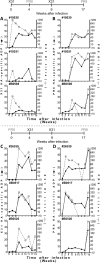
Fig 5. ADP in pigtail macaques infected with IAVs.
HI titres (grey open symbol; dotted line) and ADP activity (black filled symbol; solid line) of 6 pigtail macaques infected twice with X31 (H3N2 A/HKx31) and PR8 (H1N1 A/Puerto Rico/08/1939). Grey arrows indicate influenza strain and time of infection which is summarized above the panels–the macaques in the top panels (A, B) were first infected with X31 and macaques in the bottom panels were first infected with PR8 (C, D). X31 and PR8 immune responses for each macaque are illustrated on A, C and B, D respectively. All ADP responses were corrected subtracting influenza-naïve macaque (mean of 3 animals) background.
Fig 6
Fig 6. Influenza virus is not released from THP-1 cells after antibody-mediated virus uptake.
(A) The assay setup. Sialidase treated THP-1 cells and untreated A549 cells were infected with PR8 virus (M.O.I. of 10). Virus was previously opsonised or not with IgG from either naïve or influenza infected macaques. IgG was diluted 1:1000 to avoid neutralization of virus. Cells were washed twice to eliminate excess virus and antibodies from supernatant. After 20 hr of culture at 37°C, supernatant was transferred to MDCK cells. THP-1 pellets and resuspended A549 cells were fixed, permeabilized, labelled with anti-NP antibody and analysed by flow cytometry. 20 hr later, infection rate in MDCK was measured with intracellular NP staining. (B) Confirms antibody-mediated uptake of the PR8 virus by the THP-1 cells using sera post PR8 infection. (C) Shows the ability of supernatant from the antibody-mediated infection of THP-1 cells, or control THP-1 or A549 cells in the absence of antibody, to infect MDCK cells. Data are representative of n = 3 independent experiments.
Fig 7
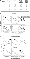
Fig 7. ADP in three humans with symptomatic H1N1 Pdm09 infection.
(A) Serologically characteristics for early and late time points after an influenza-like illness. (B) Comparison of ADP titres for the 3 subjects pre (filled symbol) and post (open symbol) influenza seroconversion of HA-coated beads from A/California/04/2009 (H1 Pdm09, squares) and A/New Caledonia/20/1999 (H1 New Cal, circles). Dotted line represents negative control ADP for an influenza naïve macaque. (C) Ability of antibodies to mediate uptake of live IAV was assessed using half log serial dilutions of IgG pre and post seroconversion (1:40 to 1:9710) as for Fig 4. Infection was compared using a matched virus strain to the infection (H1N1 Auckland Pdm09) and a mismatched strain (H1N1 PR8). Dotted line represents uptake of live virus in the absence of IgG.

References

    1. WHO. Influenza (Seasonal) 2015 [updated March 2014]. Available: http://www.who.int/mediacentre/factsheets/fs211/en/. Accessed 2015 February 7.
    1. Kilbourne ED, Smith C, Brett I, Pokorny BA, Johansson B, Cox N. The total influenza vaccine failure of 1947 revisited: major intrasubtypic antigenic change can explain failure of vaccine in a post-World War II epidemic. Proceedings of the National Academy of Sciences of the United States of America. 2002;99(16):10748–52. Epub 2002/07/24. 10.1073/pnas.162366899 ; PubMed Central PMCID: PMCPmc125033. - DOI - PMC - PubMed
    1. Tricco AC, Chit A, Soobiah C, Hallett D, Meier G, Chen MH, et al. Comparing influenza vaccine efficacy against mismatched and matched strains: a systematic review and meta-analysis. BMC medicine. 2013;11:153 Epub 2013/06/27. 10.1186/1741-7015-11-153 ; PubMed Central PMCID: PMCPmc3706345. - DOI - PMC - PubMed
    1. Talbot HK, Zhu Y, Chen Q, Williams JV, Thompson MG, Griffin MR. Effectiveness of influenza vaccine for preventing laboratory-confirmed influenza hospitalizations in adults, 2011–2012 influenza season. Clinical infectious diseases: an official publication of the Infectious Diseases Society of America. 2013;56(12):1774–7. Epub 2013/03/02. 10.1093/cid/cit124 . - DOI - PMC - PubMed
    1. Osterholm MT, Kelley NS, Sommer A, Belongia EA. Efficacy and effectiveness of influenza vaccines: a systematic review and meta-analysis. The Lancet Infectious diseases. 2012;12(1):36–44. Epub 2011/10/29. 10.1016/s1473-3099(11)70295-x . - DOI - PubMed

Publication types

MeSH terms

LinkOut - more resources