

A high fat diet alters metabolic and bioenergetic function in the brain: A magnetic resonance spectroscopy study
- PMID: 27125544
- PMCID: PMC4900919
- DOI: 10.1016/j.neuint.2016.04.008
A high fat diet alters metabolic and bioenergetic function in the brain: A magnetic resonance spectroscopy study
Abstract
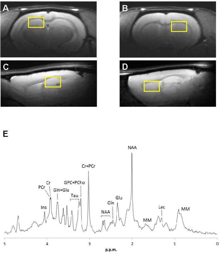
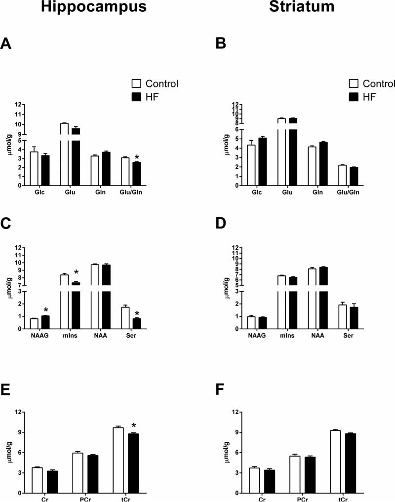
Diet-induced obesity and associated metabolic effects can lead to neurological dysfunction and increase the risk of developing Alzheimer's disease (AD) and Parkinson's disease (PD). Despite these risks, the effects of a high-fat diet on the central nervous system are not well understood. To better understand the mechanisms underlying the effects of high fat consumption on brain regions affected by AD and PD, we used proton magnetic resonance spectroscopy ((1)H-MRS) to measure neurochemicals in the hippocampus and striatum of rats fed a high fat diet vs. normal low fat chow. We detected lower concentrations of total creatine (tCr) and a lower glutamate-to-glutamine ratio in the hippocampus of high fat rats. Additional effects observed in the hippocampus of high fat rats included higher N-acetylaspartylglutamic acid (NAAG), and lower myo-inositol (mIns) and serine (Ser) concentrations. Post-mortem tissue analyses revealed lower phosphorylated AMP-activated protein kinase (pAMPK) in the striatum but not in the hippocampus of high fat rats. Hippocampal pAMPK levels correlated significantly with tCr, aspartate (Asp), phosphoethanolamine (PE), and taurine (Tau), indicating beneficial effects of AMPK activation on brain metabolic and energetic function, membrane turnover, and edema. A negative correlation between pAMPK and glucose (Glc) indicates a detrimental effect of brain Glc on cellular energy response. Overall, these changes indicate alterations in neurotransmission and in metabolic and bioenergetic function in the hippocampus and in the striatum of rats fed a high fat diet.
Keywords: Brain metabolism; Diet-induced obesity; High-fat; Imaging; Magnetic resonance spectroscopy.
Copyright © 2016 Elsevier Ltd. All rights reserved.
Figures

Similar articles
-
High-fat diet-induced hyperglutamatergic activation of the hippocampus in mice: A proton magnetic resonance spectroscopy study at 9.4T.Neurochem Int. 2018 Mar;114:10-17. doi: 10.1016/j.neuint.2017.12.007. Epub 2017 Dec 21. Neurochem Int. 2018. PMID: 29274351
-
Region-specific differences in bioenergetic proteins and protein response to acute high fat diet in brains of low and high capacity runner rats.Neurosci Lett. 2018 May 1;674:49-53. doi: 10.1016/j.neulet.2018.03.009. Epub 2018 Mar 6. Neurosci Lett. 2018. PMID: 29522838 Free PMC article.
-
Spatio-temporal metabolic rewiring in the brain of TgF344-AD rat model of Alzheimer's disease.Sci Rep. 2022 Oct 10;12(1):16958. doi: 10.1038/s41598-022-20962-6. Sci Rep. 2022. PMID: 36216838 Free PMC article.
-
Hyperpolarized [1-13C] pyruvate MR spectroscopy detect altered glycolysis in the brain of a cognitively impaired mouse model fed high-fat diet.Mol Brain. 2018 Dec 18;11(1):74. doi: 10.1186/s13041-018-0415-2. Mol Brain. 2018. PMID: 30563553 Free PMC article.
-
Region-specific cerebral metabolic alterations in streptozotocin-induced type 1 diabetic rats: an in vivo proton magnetic resonance spectroscopy study.J Cereb Blood Flow Metab. 2015 Nov;35(11):1738-45. doi: 10.1038/jcbfm.2015.111. Epub 2015 Jun 3. J Cereb Blood Flow Metab. 2015. PMID: 26036938 Free PMC article.
Cited by
-
Effect of berrycactus fruit (Myrtillocactus geometrizans) on glutamate, glutamine, and GABA levels in the frontal cortex of rats fed with a high-fat diet.Open Life Sci. 2023 Jan 24;18(1):20220529. doi: 10.1515/biol-2022-0529. eCollection 2023. Open Life Sci. 2023. PMID: 36742451 Free PMC article.
-
Failure of diet-induced transcriptional adaptations in alpha-synuclein transgenic mice.Hum Mol Genet. 2023 Jan 13;32(3):450-461. doi: 10.1093/hmg/ddac205. Hum Mol Genet. 2023. PMID: 36001352 Free PMC article.
-
Docosahexaenoic acid enhances hippocampal insulin sensitivity to promote cognitive function of aged rats on a high-fat diet.J Adv Res. 2023 Mar;45:31-42. doi: 10.1016/j.jare.2022.04.015. Epub 2022 Apr 30. J Adv Res. 2023. PMID: 35618634 Free PMC article.
-
Overweight and diabetes prevention: is a low-carbohydrate-high-fat diet recommendable?Eur J Nutr. 2018 Jun;57(4):1301-1312. doi: 10.1007/s00394-018-1636-y. Epub 2018 Mar 14. Eur J Nutr. 2018. PMID: 29541907 Free PMC article. Review.
-
Neurochemical Modifications in the Hippocampus, Cortex and Hypothalamus of Mice Exposed to Long-Term High-Fat Diet.Front Neurosci. 2019 Jan 8;12:985. doi: 10.3389/fnins.2018.00985. eCollection 2018. Front Neurosci. 2019. PMID: 30670942 Free PMC article.
References
-
- Marshall JA, Bessesen DH. Dietary fat and the development of type 2 diabetes. Diabetes Care. 2002;25(3):620–622. - PubMed
-
- Lindqvist A, Mohapel P, Bouter B, Pizzo D, Brundin P, Erlanson-Albertsson C. High-fat diet impairs hippocampal neurogenesis in male rats. Eur J Neurol. 2006;13(12):1385–1388. - PubMed
-
- Pipatpiboon N, Pratchayasakul W, Chattipakorn N, Chattipakorn SC. PPARgamma agonist improves neuronal insulin receptor function in hippocampus and brain mitochondria function in rats with insulin resistance induced by long term high-fat diets. Endocrinology. 2012;153(1):329–338. - PubMed
Publication types
MeSH terms
Grants and funding
LinkOut - more resources
Full Text Sources
Other Literature Sources
