Review
. 2016 Jun;12(6):358-68.
doi: 10.1038/nrneurol.2016.59. Epub 2016 Apr 29.

Advanced MRI and staging of multiple sclerosis lesions

Affiliations
Review

Advanced MRI and staging of multiple sclerosis lesions

Martina Absinta et al. Nat Rev Neurol. 2016 Jun.

Abstract

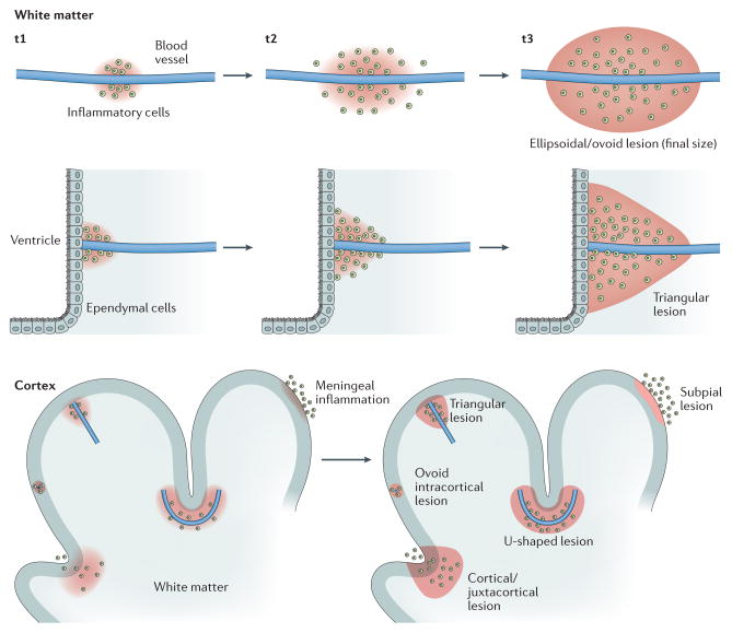
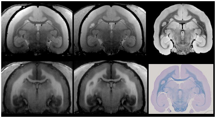
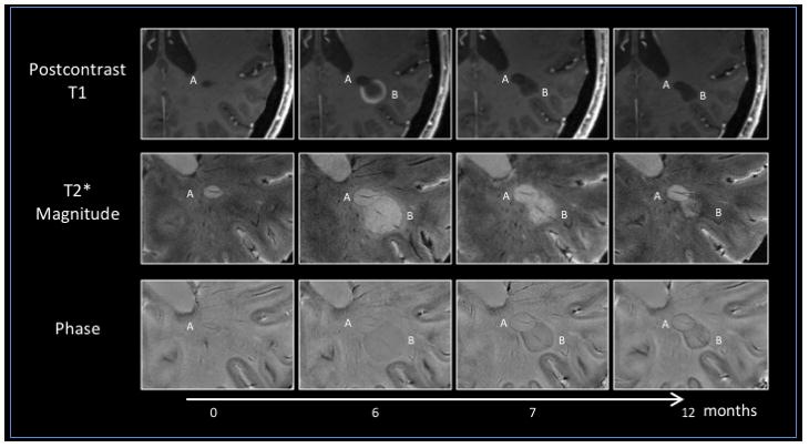
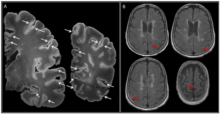
Over the past few decades, MRI-based visualization of demyelinated CNS lesions has become pivotal to the diagnosis and monitoring of multiple sclerosis (MS). In this Review, we outline current efforts to correlate imaging findings with the pathology of lesion development in MS, and the pitfalls that are being encountered in this research. Multimodal imaging at high and ultra-high magnetic field strengths is yielding biologically relevant insights into the pathophysiology of blood-brain barrier dynamics and both active and chronic inflammation, as well as mechanisms of lesion healing and remyelination. Here, we parallel the results in humans with advances in imaging of a primate model of MS - experimental autoimmune encephalomyelitis (EAE) in the common marmoset - in which demyelinated lesions resemble their human counterparts far more closely than do EAE lesions in the rodent. This approach holds promise for the identification of innovative biological markers, and for next-generation clinical trials that will focus more on tissue protection and repair.


Conflict of interest statement

statement The authors declare no competing interests.

Figures

Figure 1
Figure 1. MRI and histology of the marmoset brain
A common marmoset housed at the NIH Intramural Research Program facility was studied in accordance with the standards of the American Association for Accreditation of Laboratory Animal Care and the National Institute of Neurological Disorders and Stroke’s Animal Care and Use Committee. a,b | Baseline 7 T MRI showing a healthy brain on proton density-weighted (a) and T1-weighted (b) images. After MRI, the animal was immunized with 200 μg of fresh-frozen human white matter homogenate, and started developing white matter lesions soon after. c,d | These lesions are visible on follow-up proton density-weighted (c) and T1 weighted (d) MRI scans performed before termination of the experiment. Note that a central vein within the lesion (red arrow) can be discerned on the proton density image (c). e,f | 7 T post-mortem 100 μm isotropic MRI after necropsy (e) was used to guide tissue processing for histology (f), allowing precise correlation between MRI and histopathology (red arrows). Complete demyelination, as seen on Luxol fast blue staining (f), matches the radiological appearance of the lesion both in vivo and post mortem (c, d, e).
Figure 2
Figure 2. Perivenular topography of demyelinated MS lesions in white and grey matter
In the newly forming MS lesion, the distribution of myelin-scavenging inflammatory cells, spreading along and outward from an inflamed central vein, largely dictates the final lesion configuration. An ellipsoidal and/or rounded configuration is typical of MS lesions in the white matter. Of note, the shape of MS lesions can be also affected by the macrostructure of the surrounding tissue. For example, both the ependymal wall of the ventricles and the cortex may act as barriers to the spread of myelin-scavenging inflammatory cells. Depending of the trajectory of the vein and its position relative to these structures, lesions might assume a triangular shape, for periventricular and leukocortical–juxtacortical lesions, or a U shape, for leukocortical–juxtacortical lesions. The lower panel shows the various types of cortical demyelination. In addition to vasculocentric demyelination, which occurs in purely intracortical and leukocortical lesions, plaque-like demyelination can affect the subpial layers of the cortex and is thought to be directly triggered by leptomeningeal inflammation. MS, multiple sclerosis; t, time point.
Figure 3
Figure 3. Serial MRI evaluations in progressive MS
An untreated 49-year-old woman with progressive MS and radiological relapses underwent serial MRI evaluations at 7 T under an institutional review board-approved natural history protocol at the NIH. Lesion A is a pre-existing, non-enhancing lesion with a paramagnetic rim visible on both T2*-weighted magnitude and phase images, unknown onset, lesion A). Lesion B, a new centripetally enhancing MS lesion (open ring on postcontrast T1-weighted images; Supplementary information S2 (movie)), appears adjacent to lesion A at month 6. Both lesions exhibit a central vein on T2*-weighted magnitude and phase images. In lesion B, the peripheral T2*-weighted magnitude and phase rim persists after resolution of contrast enhancement (1 and 6 months after lesion onset). Of note, lesion B shrinks over time after enhancement resolves, whereas lesion A nearly triples in size, even in the absence of visible contrast enhancement on postcontrast T1-weighted images. In both lesions by month 12, the persistent T2*-weighted magnitude and phase rim has become darker and thicker, suggesting accrual of paramagnetic substances at the lesion edge. Pathologically, the rim possibly results from macrophages loaded with iron, and lesions with this radiological feature might, therefore, correspond to the so-called ‘chronic active’ or ‘smouldering’ lesions. MS, multiple sclerosis.
Figure 4
Figure 4. Cortical and leptomeningeal lesions
a | 7 T post-mortem MRI (two representative coronal slices of the left hemisphere, T2*-weighted gradient-echo sequence, 64 nl isotropic voxels) in a 66-year-old woman with progressive multiple sclerosis and disease duration (time from symptom onset to death) of 20 years. The arrows indicate widespread involvement of the cortex in the demyelination process, including subpial cortical and leukocortical lesions. Cortical lesions often face each other across sulci, consistent with the idea that leptomeningeal inflammation has a direct pathogenic role in cortical lesion development. b | Several foci of leptomeningeal contrast enhancement (arrows) were apparent in this patient on postcontrast 3 T MRI in vivo.

References

    1. Filippi M, et al. Association between pathological and MRI findings in multiple sclerosis. Lancet Neurol. 2012;11:349–360. - PubMed
    1. ‘t Hart BA, van Kooyk Y, Geurts JJ, Gran B. The primate autoimmune encephalomyelitis model; a bridge between mouse and man. Ann Clin Transl Neurol. 2015;2:581–593. - PMC - PubMed
    1. Maggi P, et al. The formation of inflammatory demyelinated lesions in cerebral white matter. Ann Neurol. 2014;76:594–608. - PMC - PubMed
    1. Guy JR, et al. Custom fit 3D-printed brain holders for comparison of histology with MRI in marmosets. J Neurosci Methods. 2016;257:55–63. - PMC - PubMed
    1. Ransohoff RM, Engelhardt B. The anatomical and cellular basis of immune surveillance in the central nervous system. Nat Rev Immunol. 2012;12:623–635. - PubMed

MeSH terms