. 2016 Jun;5(1):101-8.
doi: 10.1007/s40119-016-0060-8. Epub 2016 Apr 28.

Identification and Localization of Gold Nanoparticles in Potassium Ion Pores: Implications for Kir Blockade

Affiliations

Identification and Localization of Gold Nanoparticles in Potassium Ion Pores: Implications for Kir Blockade

Chur Chin et al. Cardiol Ther. 2016 Jun.

Abstract

Introduction: In our previous study, we found that negatively charged gold nanoparticles with spermidine have the potential of blocking inwardly rectifying potassium channels (Kir), both at the cellular and the tissue level.

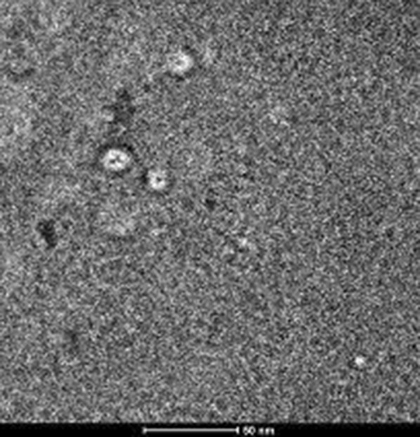
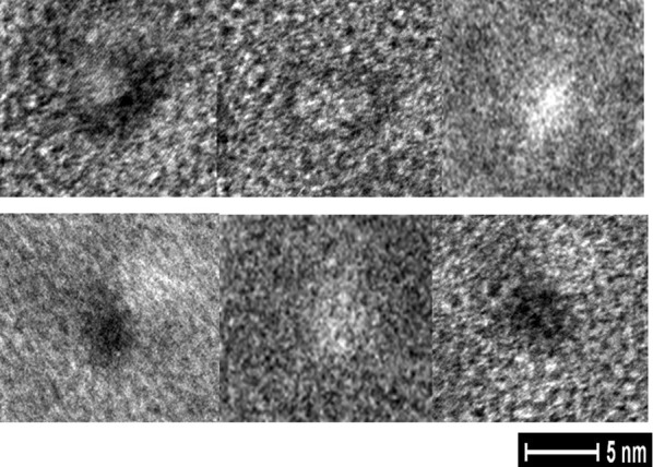
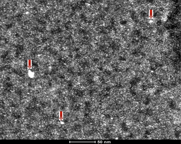
Methods: For the purpose of the present study, we purified the cytoplasmic domain of the Kir 3.1 receptor from Escherichia coli. Using single particles with surface coating by transmission electron microscopy, we identified the gold nanoparticles at the cytoplasmic side of the human Kir channel.

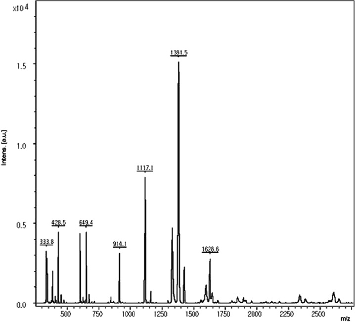
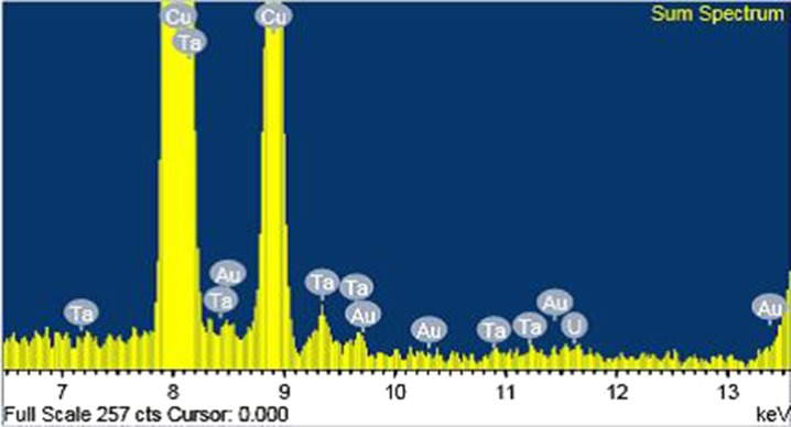
Results: Energy-dispersive X-ray spectroscopy showed the presence of the gold deposits in the cytoplasmic domain of the Kir receptor.

Conclusion: In conclusion, we could identify undecagold in the ion pore of the Kir3.1 channel in order to clarify its direct blocking effect in the Kir ion pore by undecagold.

Keywords: CryoEM; Cytoplasmic domain; Kir3.1; STEM.


Figures

Fig. 1
Fig. 1
Control (Kir receptor cytoplasmic domain): a purified protein of the cytoplsmic domain of the Kir channel have collapsed lumens. K ir inward-rectifier potassium ion channel
Fig. 2
Fig. 2
Two dimensional TEM images show the tetramer of Kir3.1 cytoplasmic domain with collapsed lumen of the ion pore. TEM transmission electron microscopy, K ir inward-rectifier potassium ion channel
Fig. 3
Fig. 3
Mass spectra of the tetramer of Kir3.1 cytoplasmic domain from the MALDI set-up shows the peptide fragments of the protein. K ir inward-rectifier potassium ion channel, MALDI matrix-assisted laser desorption/ionization
Fig. 4
Fig. 4
Treated by gold nanoparticles: single particle analysis with cryoEM shows the cytoplasmic Kir domain containing undeca-gold inside the complex
Fig. 5
Fig. 5
Two-dimensional TEM images show the tetramer of Kir3.1 cytoplasmic domain with dilated lumen of the ion pore by gold nanoparticles. TEM transmission electron microscopy, K ir inward-rectifier potassium ion channel
Fig. 6
Fig. 6
Mass spectra of tetramer of Kir3.1 cytoplasmic domain from the MALDI set-up shows the peptide fragments of the protein containing gold peaks (red arrow). K ir inward-rectifier potassium ion channel, MALDI matrix-assisted laser desorption/ionization
Fig. 7
Fig. 7
Treated by gold nanoparticles coating with DMSA-spermidine complex. DMSA meso-2.3-dimercaptosuccinic acid
Fig. 8
Fig. 8
Two-dimensional TEM images of tetramer of Kir3.1 cytoplasmic domain after treating with undecagold cating with DMSA—spermidine complex shows the hyperdense DMSA coating in the ion pore or adjacent tetramer. TEM transmission electron microscopy, K ir inward-rectifier potassium ion channel, DMSA meso-2.3-dimercaptosuccinic acid
Fig. 9
Fig. 9
EDS by STEM of the cytoplasmic domain of the receptor treated by gold nanoparticles: transmission EM (TEM) with surface-coated with DMSA have been successfully used to produce a high signal in TEM images in the ion pore or surrounding tetramer components. EDS energy dispersive X-ray spectroscopy, STEM scanning transmission electron microscopy
Fig. 10
Fig. 10
Two-dimensional STEM images for the EDS shows hyperdense gold nanoparticle (red arrow) in the lumens of the tetramer of the cytoplasmic domain. STEM scanning transmission electron microscopy, EDS energy dispersive X-ray spectroscopy
Fig. 11
Fig. 11
The EDS spectrum of the receptors containing the gold nanoparticles in it indicates the presence of C and O from the receptor molecules. The small gold peak evident in the EDS spectrum confirms that a validated amount of the 0.8 nm gold nanoparticle is present in the ion pore of Kir channels. EDS energy dispersive X-ray spectroscopy, K ir inward-rectifier potassium ion channel

References

    1. Nicholas CG, Makhina EN, Pearson WL, Sha Q, Lopatin AN. Inward rectification and implications for cardiac excitability. Circ Res. 1996;78:1–7. doi: 10.1161/01.RES.78.1.1. - DOI - PubMed
    1. Wilczewska AZ, Niemirowicz K, Markiewicz KH, Car H. Nanoparticles as drug delivery systems. Pharmacol Rep. 2012;64:1020–1037. doi: 10.1016/S1734-1140(12)70901-5. - DOI - PubMed
    1. Chin C, Kim IK, Lim DY, Kim KS, Lee HA, et al. Gold nanoparticls-choline complexes can block nicotinic acetylcholine receptors. Int J Nanomed. 2010;5:315–321. doi: 10.2147/IJN.S10466. - DOI - PMC - PubMed
    1. Leifert A, Pan Y, Kinkeldey A, Schiefer F, Setzler J, Scheel O, et al. Differential hERG ion channel activity of ultrasmall gold nanoparticles. PNAS. 2013;110:8004–8009. doi: 10.1073/pnas.1220143110. - DOI - PMC - PubMed
    1. Chin C. Gold nanoparticle-spermidine complex blocks the inward rectifier potassium channel. Am J Cardiovasc Dis. 2014;4:34–46. - PMC - PubMed

LinkOut - more resources