. 2016;17(1):35-45.
doi: 10.3727/105221616X691578. Epub 2016 Apr 27.

Liver-Specific Deletion of Integrin-Linked Kinase in Mice Attenuates Hepatotoxicity and Improves Liver Regeneration After Acetaminophen Overdose

Affiliations

Liver-Specific Deletion of Integrin-Linked Kinase in Mice Attenuates Hepatotoxicity and Improves Liver Regeneration After Acetaminophen Overdose

Bharat Bhushan et al. Gene Expr. 2016.

Abstract

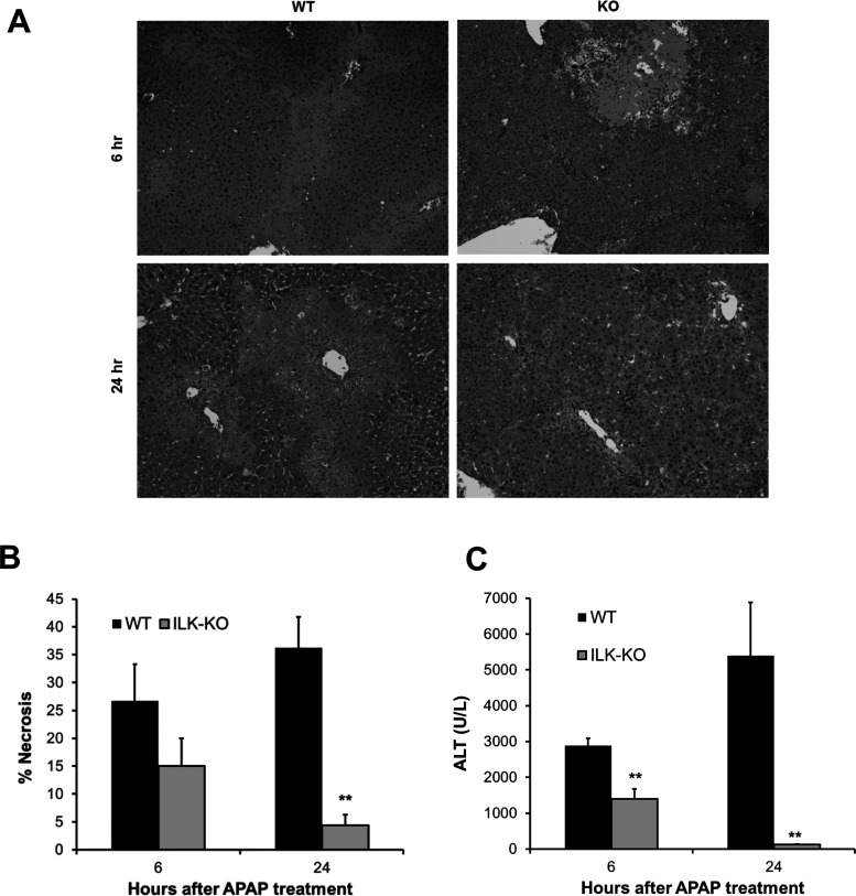
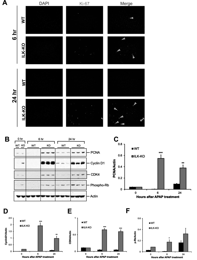
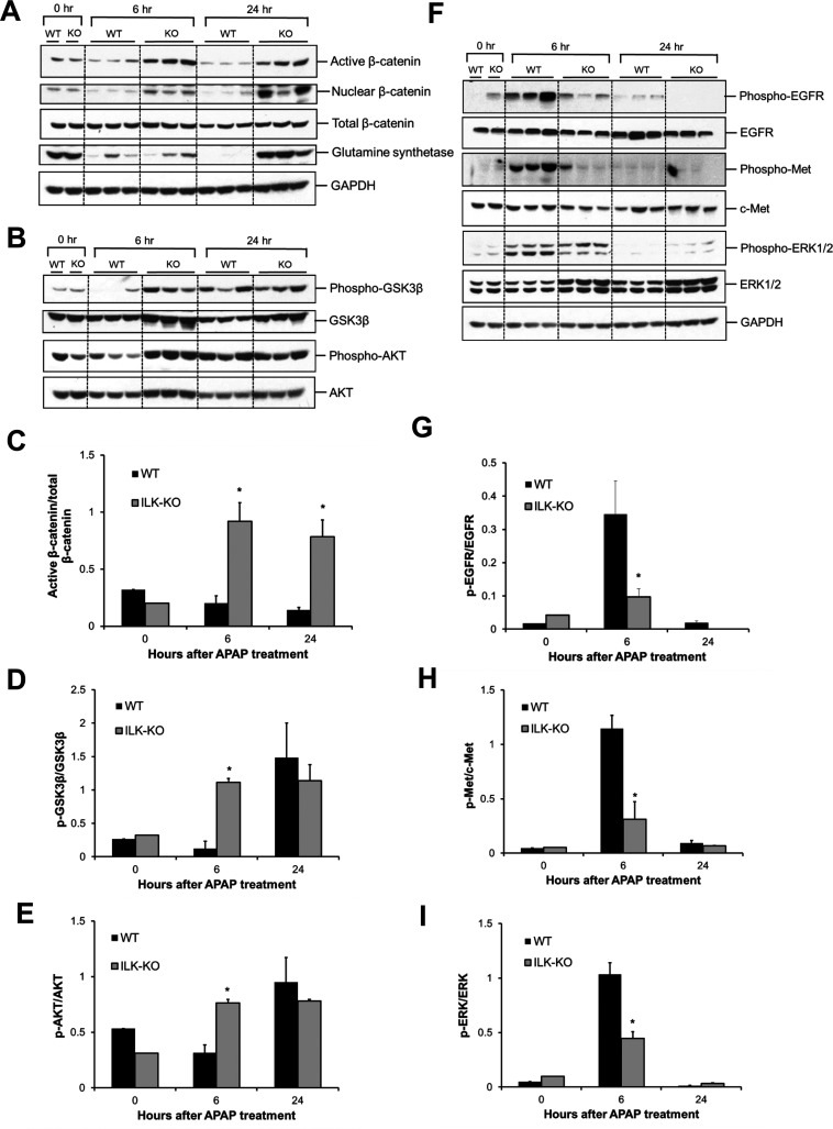
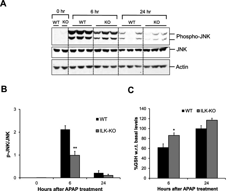
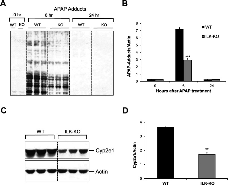
Acetaminophen (APAP) overdose is the major cause of acute liver failure in the US. Prompt liver regeneration is critical for recovery after APAP hepatotoxicity, but mechanisms remain elusive. Extracellular matrix (ECM)-mediated signaling via integrin-linked kinase (ILK) regulates liver regeneration after surgical resection. However, the role of ECM signaling via ILK in APAP toxicity and compensatory regeneration is unknown, which was investigated in this study using liver-specific ILK knockout (KO) mice. ILK KO and wild-type (WT) mice were treated with 300 mg/kg APAP, and injury and regeneration were studied at 6 and 24 h after APAP treatment. ILK KO mice developed lower liver injury after APAP overdose, which was associated with decreased JNK activation (a key mediator of APAP toxicity). Further, higher glutathione levels after APAP treatment and lower APAP protein adducts levels, along with lower levels of CYP2E1, suggest decreased metabolic activation of APAP in ILK KO mice. Interestingly, despite lower injury, ILK KO mice had rapid and higher liver regeneration after APAP overdose accompanied with increased β-catenin signaling. In conclusion, liver-specific deletion of ILK improved regeneration, attenuated toxicity after APAP overdose, and decreased metabolic activation of APAP. Our study also indicates that ILK-mediated ECM signaling plays a role in the regulation of CYP2E1 and may affect toxicity of several centrilobular hepatotoxicants including APAP.


Figures

Figure 1
Figure 1
Attenuated liver injury after acetaminophen (APAP) overdose in integrin-linked kinase (ILK) knockout (KO) mice. (A) Representative photomicrographs of hematoxylin and eosin (H&E)-stained liver sections. (B) Bar graph showing percent necrosis area based on H&E-stained liver sections. (C) Bar graph showing serum aminotransferase (ALT) levels. All samples were collected from wild-type (WT) or ILK KO mice treated with 300 mg/kg APAP. Significant difference between groups at **p < 0.01.
Figure 2
Figure 2
Improved liver regeneration after APAP overdose in ILK KO mice. (A) Representative photomicrographs showing Ki-67 immunofluorescence performed on frozen liver sections of WT or ILK KO mice treated with 300 mg/kg APAP. (B) Western blot analysis of proliferating cell nuclear antigen (PCNA), cyclin D1, CDK4, and phospho-Rb using total cell extract of liver of WT or ILK KO mice treated with 300 mg/kg APAP. All samples were collected at 0, 6, and 24 h after APAP treatment. Densitometric analysis showing quantification of protein expression of (C) PCNA, (D) cyclin D1, (E) CDK4, and (F) phospho-Rb relative to actin as loading control. Significant difference between groups at *p < 0.05, **p < 0.01, and ***p < 0.001.
Figure 3
Figure 3
Differential activation of proregenerative signaling after APAP overdose in ILK KO mice. (A) Western blot analysis of active β-catenin, nuclear β-catenin, total β-catenin, glutamine synthetase, (B) phospho-GSK3β, GSK3β, phospho-AKT, and AKT using total cell extract (unless specified) of liver of WT or ILK KO mice treated with 300 mg/kg APAP. Densitometric analysis showing (C) activation of β-catenin, (D) inactivation of GSK3β, and (E) activation of AKT, respectively, based on Western blot images shown in (A) and (B). (F) Western blot analysis of phospho-EGFR, EGFR, phospho-Met, c-Met, phospho-ERK1/2, and ERK1/2 using total cell extract of liver of WT or ILK KO mice treated with 300 mg/kg APAP. (G–I) Densitometric analysis showing quantification of expression proteins shown in (F). All samples were collected at 0, 6, and 24 h after APAP treatment. Significant difference between groups at *p < 0.05.
Figure 4
Figure 4
Attenuated injury signaling in liver of ILK KO mice after APAP overdose. (A) Western blot analysis of phospho-JNK and JNK using total cell extract of liver of WT or ILK KO mice treated with 300 mg/kg APAP. All samples were collected at 0, 6, and 24 h after APAP treatment. (B) Densitometric analysis showing quantification of protein expression of phospho-JNK relative to JNK. (C) Liver glutathione levels represented as percentage of basal levels in WT or ILK KO mice. Significant difference between groups at *p < 0.05 and **p < 0.01.
Figure 5
Figure 5
Altered APAP metabolic activation in ILK KO mice. (A) Western blot analysis showing APAP protein adduct levels in liver of WT or ILK KO mice treated with 300 mg/kg APAP. All samples were collected at 0, 6, and 24 h after APAP treatment. (B) Densitometric analysis showing quantification of APAP protein adduct levels shown in (A). (C) Western blot analysis showing CYP2E1 protein expression in the liver of WT or ILK KO mice at 0 h. (D) Densitometric analysis showing quantification of CYP2E1 levels shown in (C). Significant difference between groups at **p < 0.01 and ***p < 0.001.

References

    1. Bernal W, Lee WM, Wendon J, Larsen FS, Williams R. Acute liver failure: A curable disease by 2024? J Hepatol. 2015;62(1S):S112–20. - PubMed
    1. Jaeschke H, McGill MR, Ramachandran A. Oxidant stress, mitochondria, and cell death mechanisms in drug-induced liver injury: Lessons learned from acetaminophen hepatotoxicity. Drug Metab Rev. 2012;44(1):88–106. - PMC - PubMed
    1. Bhushan B, Walesky C, Manley M, Gallagher T, Borude P, Edwards G, Monga SP, Apte U. Pro-regenerative signaling after acetaminophen-induced acute liver injury in mice identified using a novel incremental dose model. Am J Pathol. 2014;184(11):3013–25. - PMC - PubMed
    1. Donahower BC, McCullough SS, Hennings L, Simpson PM, Stowe CD, Saad AG, Kurten RC, Hinson JA, James LP. Human recombinant vascular endothelial growth factor reduces necrosis and enhances hepatocyte regeneration in a mouse model of acetaminophen toxicity. J Pharmacol Exp Ther. 2010;334(1):33–43. - PMC - PubMed
    1. Hu B, Colletti LM. Stem cell factor and c-kit are involved in hepatic recovery after acetaminophen-induced liver injury in mice. Am J Physiol Gastrointest Liver Physiol. 2008;295(1):G45–53. - PMC - PubMed

MeSH terms