. 2016 Apr 29;352(6285):550-5.
doi: 10.1126/science.aad4821.

Changes in the composition of brain interstitial ions control the sleep-wake cycle

Affiliations

Changes in the composition of brain interstitial ions control the sleep-wake cycle

Fengfei Ding et al. Science. .

Abstract

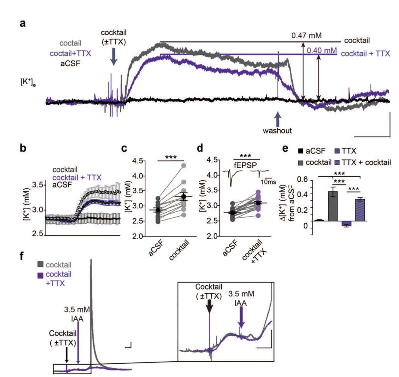
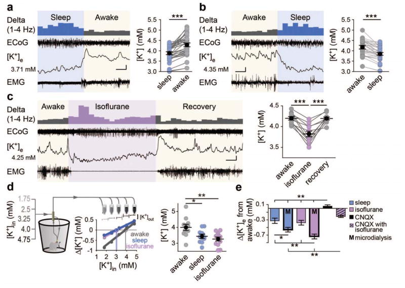
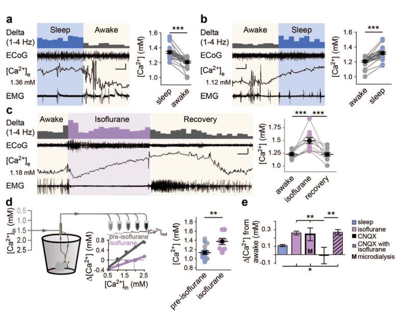
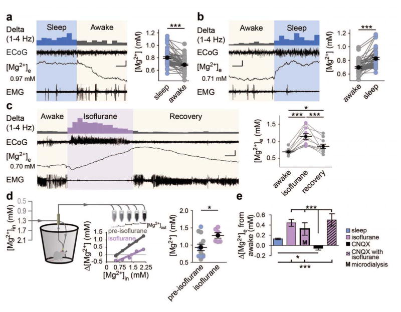
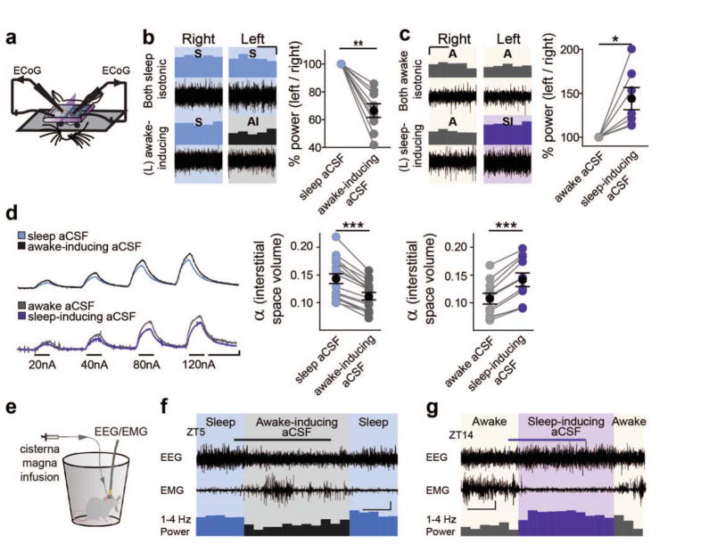
Wakefulness is driven by the widespread release of neuromodulators by the ascending arousal system. Yet, it is unclear how these substances orchestrate state-dependent, global changes in neuronal activity. Here, we show that neuromodulators induce increases in the extracellular K(+) concentration ([K(+)]e) in cortical slices electrically silenced by tetrodotoxin. In vivo, arousal was linked to AMPA receptor-independent elevations of [K(+)]e concomitant with decreases in [Ca(2+)]e, [Mg(2+)]e, [H(+)]e, and the extracellular volume. Opposite, natural sleep and anesthesia reduced [K(+)]e while increasing [Ca(2+)]e, [Mg(2+)]e, and [H(+)]e as well as the extracellular volume. Local cortical activity of sleeping mice could be readily converted to the stereotypical electroencephalography pattern of wakefulness by simply imposing a change in the extracellular ion composition. Thus, extracellular ions control the state-dependent patterns of neural activity.


Figures

Fig. 1
Fig. 1
Neuromodulators increase [K+]e concentration in the absence of neuronal activity. (A) Representative traces of [K+]e shifts before, during, and after administering the neuromodulator cocktail. Scale bar: x = 5 min, y = 0.2 mM [K+]e (B) 30 s binned averages of shifts before and after neuromodulator cocktail (C and D) Summary of [K+]e increase following neuromodulator cocktail application in slices ± TTX (paired t-test, C (-TTX): t(11) = 5.871, P = 0.0001; D (+TTX), t(21) = 11.69, P < 0.0001). Inset: representative fEPSP recordings before and after application of TTX. Scale bar = 10 ms (E) Summarized shifts at 10 minutes following changing perfusion solution. n = 6 (cocktail-free aCSF), 12 (cocktail), 10 (aCSF+TTX), 22 (cocktail+TTX). One-way ANOVA: F(3,46) = 25.94, P < 0.0001. Post-hoc Tukey test: ** P < 0.01, ***P < 0.001. (F) Representative trace of large [K+]e spike following metabolic stress using iodoacetate (IA). Trace includes pre-cocktail baseline, aCSF + cocktail, aCSF + IA and return to baseline aCSF. (Right) Magnified trace showing [K+]e increases in slices treated with the neuromodulator cocktail (±TTX) followed by IA. Scale bars: x = 5 min, y = 0.2 mM [K+]e. Mean (black circle) ± SEM.
Fig. 2
Fig. 2
Extracellular K+ is higher during wakefulness. (A and B) Representative ECoG, [K+]e, and EMG recordings with a data summary of state transitions in sleep to awake (A) or awake to sleep (B) Initial [K+]e concentrations are shown to the left, and the 1–4 Hz power is displayed above to illustrate state-dependent shifts, binned at 10 s for clarity. n = 34 transitions; One-way, repeated measures ANOVA: F(3,99) = 9.536, P < 0.0001. Tukey post-hoc multiple comparisons test: ***P < 0.001. Scale bar: x = 20 s, y = 0.1 mM [K+]e; ECoG = 0.75 mV; EMG = 0.3 mV (A), 1 mV (B). (C) Representative recording of awake to isoflurane transitions recorded during the natural awake period (ZT16-20). n = 11 animals. One-way repeated measures ANOVA: F(2,20) = 35.61, P < 0.0001. Post-hoc Tukey test: ***P < 0.001. Scale bar: x = 1.5 min, y = 0.15 mM [K+]e, 0.75 mV EMG and ECoG. (D) Microdialysis samples collected from freely moving mice during their awake (ZT14-20) or sleeping (ZT2-8) period, and under isoflurane anesthesia. (Left) Schematic illustrating setup and inflow [K+] gradient with representative ISM trace and sample no-net flux method plot used for [K+]e estimate. (right) Summary of [K+]e by state: n = 12 awake, 12 sleep, and 13 isoflurane animals. One-way ANOVA: F(2, 34) = 8.055, P = 0.0014. Post-hoc Tukey test: *P <0.05, **P < 0.01. (E) Comparison of all state-dependent transitions. One-way ANOVA comparing mean shifts by group: F(5,161) = 16.61, P < 0.0001. Post-hoc Tukey test: *P < 0.05, **P < 0.01. Mean (black circle) ± SEM
Fig. 3
Fig. 3
Extracellular Ca2+ decreases during wakefulness. (A and B) Representative ECoG, EMG, and [Ca2+]e recordings in sleep to awake (A) and awake to sleep (B). Initial [Ca2+]e is listed to the left with 1–4 Hz power presented above. n = 28 transitions. One-way, repeated measures ANOVA: F(3,81) = 39.91, P < 0.0001. Post-hoc Tukey test: ***P < 0.001. Scale bars: x = 20 s (A), 40 s (B), y: [Ca2+]e = 0.05 mM, 0.75 mV EMG/ECoG (C) Representative recording of isoflurane induction and recovery (ZT16-20) and data summary. n = 11 animals. One-way repeated measures ANOVA: F(2,20) = 18.52, P < 0.0001. Post-hoc Tukey test: ***P < 0.001. Scale bar: x = 6 min, y = [Ca2+]e = 0.2 mM, 0.5 mV EMG/ECoG. (D) Schematic of microdialysis collection. Individual sleep (light blue) and awake (gray) data are pooled and compared to isoflurane. n = 8 awake, 8 sleep, and 8 isoflurane. Two-tailed t-test of isoflurane versus non isoflurane: t(22) = 3.420, P = 0.003. **P < 0.01. (E) Comparisons of [Ca2+]e shifts from the awake to sleep, isoflurane, CNQX and CNQX + isoflurane. n =8 animals CNQX and CNQX + Isoflurane One-way ANOVA comparison of state-dependent shifts: F(4,117) = 5.824, P = 0.0003; Post-hoc Tukey test: *P < 0.05, **P < 0.01. Mean (black circle) ± SEM.
Fig. 4
Fig. 4
Extracellular Mg2+ decreases during wakefulness. (A and B) Representative state-transitions between sleep and awake ECoG, EMG, and Mg2+-sensitive microelectrodes with data summary. Initial [Mg2+]e is given to the left of the ISM trace. n = 73 sleep to awake transitions (A) and 58 awake to sleep transitions (B). One-way ANOVA: F(3, 258) = 28.13, P < 0.0001. Post-hoc Tukey test: ***P < 0.001. Scale bars: x = 20 s, y = 0.05 mM [Mg2+]e, 0.33 mV EMG/ECoG. (C) Representative awake to isoflurane recording (ZT16-20) and summary of changes. n = 11 animals. One-way repeated measures ANOVA: F(2,20) = 28.13, P < 0.0001. Post-hoc Tukey test: *P < 0.05, ***P < 0.001. Scale bar: x = 5 minutes, y = 0.25 mM [Mg2+]e, 0.6 mV EMG/ECoG. (D) Schematic of microdialysis collection with data summary. Representative Mg2+-ISM and no-net flux calculation are shown. Sleep (light blue) and awake (gray) are pooled and compared to isoflurane. n = 8 awake, 8 sleep, and 8 isoflurane. Two-tailed t-test of isoflurane versus pre-isoflurane: t(16) = 2.427, P = 0.0274. (*P < 0.05). (E) Comparison of state-dependent [Mg2+]e shifts from awake to sleep, isoflurane, CNQX and awake + CNQX to isoflurane + CNQX. n = awake to CNQX: 6 animals; awake + CNQX to isoflurane + CNQX: 6 animals. One-way ANOVA of relative state-dependent shifts: F(4,167) = 27.31, P < 0.0001. Post-hoc Tukey test: **P < 0.01, ***P < 0.001. Mean (black circle) ± SEM.
Fig. 5
Fig. 5
Imposing changes in extracellular ion concentrations alter local activity, extracellular space, and behavioral state. (A) Schematic of double-cranial window recording setup. Symmetrically positioned, separate cranial windows over opposing somatosensory cortices were prepared with ECoGs simultaneously recorded in both hemispheres. (B and C) Representative ECoG recordings in B sleeping (ZT4-8), and C awake (ZT16-20) mice. (top) Representative recordings in aCSF mimicking natural state-dependent interstitial ion composition. (bottom) Recordings following change of left hemisphere aCSF to awake-inducing (B) or sleep-inducing aCSF (C) (right) Summary of 1–4 Hz ECoG power shift in the left hemisphere, normalized to the right, following change to awake-inducing (B) or sleep-inducing (C) aCSF. (paired t-test of 1–4 Hz power shifts: A: t(8) = 3.530, P = 0.008; B: t(6) = 3.091, P = 0.0214)). *P < 0.05, **P < 0.01. Scale bar: x = 4 min, y = 10% (D) TMA+ traces of shifts in extracellular space volume (α) following switch from sleep to awake-inducing aCSF (upper trace) or awake to sleep-inducing aCSF (lower trace) Note: higher amplitude = decreased dilution of TMA+, and smaller extracellular space. Data are summarized to the right. Paired t-test (awake-inducing: n = 16 animals; t(15) = 11.04, P < 0.0001; sleep-inducing: n = 11 animals; t(10) = 8.95, P < 0.0001 ) **P < 0.01. Scale: x = 2 min, y = 2 mV. (E) Schematic of cisterna-magna infusion and wire EEG/EMG recording setup. (F and G) Representative traces showing EEG and EMG activity prior, during, and following a 0.3–0.5 μl min−1 infusion of modified awake-inducing (F) or sleep-inducing (G) aCSF into the cisterna magna. The 1–4 Hz relative power (% of 1–32 Hz) is presented in averaged 10 min bins below. Infusion was run between ZT 5.5-7 (gray bar) (F) and ZT15-16.5 (purple bar) (G), during sleep and awake periods, respectively. Scale bar: x = 30 min, y = 0.5 mV (F), 1mV (G).

Comment in

References

    1. Tononi G, Cirelli C. Sleep and the price of plasticity: from synaptic and cellular homeostasis to memory consolidation and integration. Neuron. 2014;81:12–34. - PMC - PubMed
    1. Benington JH, Heller HC. Restoration of brain energy metabolism as the function of sleep. Prog Neurobiol. 1995;45:347–360. - PubMed
    1. Hinard V, et al. Key electrophysiological, molecular, and metabolic signatures of sleep and wakefulness revealed in primary cortical cultures. J Neurosci. 2012;32:12506–12517. - PMC - PubMed
    1. Saper CB, Scammell TE, Lu J. Hypothalamic regulation of sleep and circadian rhythms. Nature. 2005;437:1257–1263. - PubMed
    1. O’Donnell J, Zeppenfeld D, McConnell E, Pena S, Nedergaard M. Norepinephrine: a neuromodulator that boosts the function of multiple cell types to optimize CNS performance. Neurochem Res. 2012;37:2496–2512. - PMC - PubMed

Publication types

MeSH terms