. 2016 Apr 28;13(1):93.
doi: 10.1186/s12974-016-0558-y.

Retinal glial responses to optic nerve crush are attenuated in Bax-deficient mice and modulated by purinergic signaling pathways

Affiliations

Retinal glial responses to optic nerve crush are attenuated in Bax-deficient mice and modulated by purinergic signaling pathways

Caitlin E Mac Nair et al. J Neuroinflammation. .

Abstract

Background: Retinal ganglion cell (RGC) soma death is a consequence of optic nerve damage, including in optic neuropathies like glaucoma. The activation of the innate immune network in the retina after nerve damage has been linked to RGC pathology. Since the eye is immune privileged, innate immune functions are the responsibility of the glia, specifically the microglia, astrocytes, and Müller cells that populate the retina. Glial activation, leading to the production of inflammatory cytokines, is a hallmark feature of retinal injury resulting from optic nerve damage and purported to elicit secondary degeneration of RGC somas.

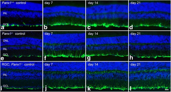
Methods: A mouse model of optic nerve crush (ONC) was used to study retinal glial activation responses. RGC apoptosis was blocked using Bax-deficient mice. Glial activation responses were monitored by quantitative PCR and immunofluorescent labeling in retinal sections of activation markers. ATP signaling pathways were interrogated using P2X receptor agonists and antagonists and Pannexin 1 (Panx1)-deficient mice with RGC-specific deletion.

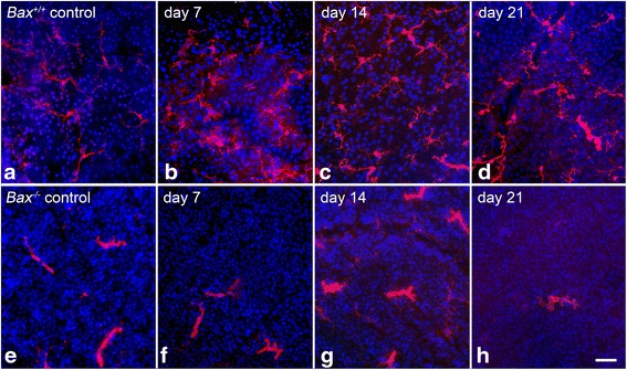
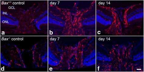
Results: ONC induced activation of both macroglia and microglia in the retina, and both these responses were dramatically muted if RGC death was blocked by deletion of the Bax gene. Macroglial, but not microglial, activation was modulated by purinergic receptor activation. Release of ATP after optic nerve damage was not mediated by PANX1 channels in RGCs.

Conclusions: RGC death in response to ONC plays a principal stimulatory role in the retinal glial activation response.

Keywords: BAX; Macroglia; Microglia; Neuroinflammation; Optic nerve damage; P2X Receptor; PANX1; Retinal ganglion cell.


Figures

Fig. 1
Fig. 1
Expression of Aif1 and Cd68 are attenuated in Bax-deficient mice after optic nerve crush. Wild-type and Bax −/− mice were subjected to crush injury and evaluated for microglial activation markers. a In wild-type mice, the mRNA expression of Aif1 peaked by 7 days post-injury, and then declined at 14 days and again at 21 days. In the Bax −/− mice, Aif1 expression was significantly attenuated relative to wild-type mice at 7 and 14 days. By 21 days, the levels of Aif1 appeared to be returning to baseline in both genotypes, and were no longer significantly different (P = 0.07). b The change in Cd68 expression followed a similar trend as Aif1, with levels peaking in the wild-type mice at 7 days before declining at 14 and again at 21 days. The Bax −/− mice, however, exhibited a significant attenuation in Cd68 expression at 7 and 14 days post-crush. By 21 days, expression in the knockout mice was significantly elevated over the wild types; however, expression in both genotypes remained low. Data is presented as mean ± SD. *P < 0.005. For each genotype at each time point, n ≥ 3
Fig. 2
Fig. 2
Upregulation of AIF1 is attenuated in Bax-deficient mice after optic nerve crush. Cross sections of eye cups were analyzed for changes in AIF1 expression after crush injury. ad Although a baseline level of AIF1-positive cells (red label) populated the retina in control eyes, following optic nerve crush Bax +/+ retinas exhibited an increase in the number of AIF1-positive cells. Microglial cells appeared in the inner plexiform layer (IPL), outer plexiform layer (IPL), and ganglion cell layer (GCL). eh In Bax −/− mice, the amount of AIF1 labeling was significantly attenuated after crush, and the intensity of AIF1 labeling did not appear to change. Inner nuclear layer (INL), outer nuclear layer (ONL). DAPI nuclear counterstain (blue label). Scale bar 50 μm. For each genotype at each time point, n ≥ 3
Fig. 3
Fig. 3
Immunofluorescent images of whole-mounted retinas stained for AIF1. Representative nerve fiber layer images of retinas from wild-type and Bax −/− mice stained for AIF1 (red label) at 7, 14, and 21 days post-crush. These are representative of the images used to quantify the number of microglia present after crush. ad In wild-type mice, a baseline resting microglial population was present in the retina. These microglia exhibited small somas with numerous long processes. After crush, the number of microglia increased, and the morphology transitioned to an amoeboid state as the cell somas thickened and the processes retracted. By 21 days, while there was still a prominent population of AIF1-positive microglia, the number of cells was beginning to decline. eh The number and morphology of microglia in the Bax −/− mice contrasted starkly with the population in the wild types. The number of microglia in the knockout mice did not increase by 7 days after crush, and after a modest increase in AIF1 labeling at 14 days, the number of microglia dropped considerably. Interestingly, the morphology of microglia in the Bax −/− mice appeared amoeboid, even in the control eyes, characteristic of an early activation state. These microglia exhibited very few processes and thickened somas. Microglial counts were obtained from at least six images, and each image is approximately 0.04 mm2. DAPI nuclear counterstain (blue label). Scale bar 50 μm. For each genotype at each time point, n ≥ 3
Fig. 4
Fig. 4
Microglial changes in the peripheral retinal nerve fiber layer of Bax −/− mice after crush. a The number of AIF1-positive cells were quantified from retinal whole mounts of wild-type and Bax −/− mice after crush. Consistent with past literature, the number of microglia in the wild-type retinas rose dramatically by 7 days after crush, and remained significantly elevated at 14 days before declining at 21 days. Microglial counts in the Bax −/− mice were significantly attenuated relative to the wild types, and although a modest increase was quantified in both the experimental and control eyes (suggesting a contralateral effect from crush injury), the number of microglia in the crushed retinas of Bax −/− mice was significantly attenuated at 7 and 14 days post-crush. By 21 days, the microglial counts were no longer statistically significant between the two genotypes. Data is presented as mean ± SE. *P < 0.0001. For each genotype at each time point, n ≥ 3. b Sholl analysis of microglial morphology showing the numbers of processes crossing the 40-μm grid line (complete data shown in Additional file 1: Figure S1) for AIF1-positive cells in both crushed and contralateral control retinas of each genotype. Wild-type microglia exhibited an increase in processes at 7 days after crush (*P < 0.0001 relative to contralateral wild-type retinas), while microglia in Bax-deficient mice exhibited a significant decrease in processes by 14 days (**P < 0.0001, relative to control retinas). By 21 days, both control and crush retinas of each genotype exhibited similar levels of ramification, which were significantly reduced compared to earlier time points (P < 0.005). Data is presented as mean ± SE. A minimum of 50 cells from at least three mice, at each time point, were measured
Fig. 5
Fig. 5
Microglial activation is present but reduced in the optic nerve head of Bax −/− mice after crush. The microglial populations in the optic nerve heads of Bax +/+ and Bax −/− mice were also compared. ac Wild-type mice exhibited a clear increase in the intensity and number of AIF1-expressing cells (red label) at both 7 and 14 days after crush. df Unlike in the retina, mice deficient for Bax also exhibited an increase in microglia at the optic nerve head 7 and 14 days post-crush, although the number of cells and intensity did not appear as high as the wild-type counterparts. DAPI nuclear counterstain (blue label). Scale bar 50 μm. For each genotype at each time point, n ≥ 3
Fig. 6
Fig. 6
Expression of macroglial markers Gfap and Nes are significantly attenuated after crush in Bax −/− mice. Wild-type and Bax −/− mice were evaluated for macroglial activation markers following optic nerve crush. a In wild-type mice, Gfap mRNA expression rose sharply by 7 days, and peaked at 14 days before declining again at 21 days post-crush. The Bax −/− mice, however, showed no change in Gfap expression 7 days after crush, and although expression was elevated by 14 days, it still remained significantly lower than the wild-type mice. By 21 days, both genotypes were trending towards baseline and were no longer significantly different. b A second macroglial activation marker, Nes, was also elevated in wild-type mice by 7 days after crush, after which expression steadily declined at 14 and again 21 days. Although Bax −/− mice also exhibited a rise in Nes expression at 7 and 14 days post-crush, the values were significantly lower than those seen in wild-type mice. By 21 days Nes expression levels were no longer significant between the wild-type and knockout mice. Data is presented as mean ± SD. *P < 0.001, **P < 0.0001. For each genotype at each time point, n ≥ 3
Fig. 7
Fig. 7
Müller cell upregulation of GFAP is absent after crush in Bax −/− mice. The expression of GFAP protein (green label) was also evaluated in retinal sections from wild-type and Bax −/− mice after crush. ad Astrocytes expressed a baseline level of GFAP in the retinas of wild-type mice, and by 7 days after crush, there was a dramatic increase in GFAP labeling in the ganglion cell layer (GCL), as well as through the retinal layers, indicative of Müller cell activation. Müller cells continued to express GFAP at 14 and 21 days, although expression levels appeared to decline at the latter time point. eh While retinas of Bax −/− mice also expressed GFAP in the astrocytes of control eyes, after crush, there was no upregulation of GFAP in the GCL, or through the retinal layers. Inner nuclear layer (INL), outer nuclear layer (ONL). DAPI nuclear counterstain (blue label). Scale bar 50 μm. For each genotype at each time point, n ≥ 3
Fig. 8
Fig. 8
NMDA treatment triggers microglial and macroglial activation in Bax −/− mice. Wild-type and Bax −/− mice received an intraocular injection of NMDA to confirm that Bax-deficient glia were capable of mounting an activation response. Mice were evaluated for microglial and macroglial activation 7 days post-injection. a Both wild-type and Bax −/− mice exhibited a significant increase in the expression of Aif1 and Gfap relative to control eyes. The values for Aif1 expression were not statistically different between the two genotypes, although Gfap expression was significantly higher in Bax −/− mice relative to the wild-type mice (*P < 0.05). b, c Immunofluorescence also revealed a clear increase in AIF1 labeling (red) in the retinas of Bax −/− mice, with microglia appearing in the ganglion cell layer (GCL) and inner and outer plexiform layers. d, e The expression of GFAP (green label) was also confirmed in Bax −/− mice. A clear increase in labeling was seen in the GCL, as well as through the retinal layers, indicating that Bax −/− Müller glia are capable of upregulating GFAP. a Data is presented as mean ± SD. Inner nuclear layer (INL), outer nuclear layer (ONL). DAPI nuclear counterstain (blue label). Scale bar 50 μm. For each genotype, n ≥ 3
Fig. 9
Fig. 9
A P2X receptor agonist triggers macroglial activation without inducing RGC injury. a Microglial activation was evaluated by monitoring Aif1 expression 48 h after an intraocular injection of BzATP. This treatment was not found to affect Aif1 mRNA levels relative to PBS-treated retinas (P = 0.21). Additionally, when mice were pre-treated with an intraocular injection of oxATP, a P2X receptor antagonist, prior to optic nerve crush, by 7 days post-crush Aif1 expression by the microglial population not statistically different from PBS-injected retinas (P = 0.20). Data is presented as mean ± SD. For each treatment group, n ≥ 3. b Wild-type mice were treated with an intraocular injection of the P2X receptor agonist, BzATP, which induced a clear increase in Gfap mRNA expression by 48 h. ce Immunofluorescence of retinal sections confirmed that BzATP triggered an increase in GFAP expression (green label), most obviously in the Müller cell population. The PBS-treated eyes did not exhibit an increase in GFAP. DAPI nuclear counterstain (blue label). Scale bar 50 μm. f The change in Gfap expression after BzATP treatment was also evaluated in Bax −/− mice. By 48 h, Gfap expression was elevated in both wild-type and knockout mice, indicative of functional purinergic receptors on Bax-deficient glia. Importantly, markers of RGC injury (Sncg and Nrn1) did not decline, indicating glial activation occurred in the absence of RGC injury. Data is presented as mean ± SD. Ganglion cell layer (GCL). *P < 0.005. For each treatment group and genotype, n ≥ 3
Fig. 10
Fig. 10
Treatment with oxATP reduces Gfap mRNA expression after crush injury. To evaluate purinergic signaling in the crush paradigm, wild-type mice were treated with oxATP either prior to or after optic nerve crush, and evaluated for macroglial expression of Gfap 7 days after injury. a Treatment with oxATP 1 day prior to optic nerve crush reduced Gfap transcript levels by about 50 %, relative to the PBS-injected eyes. bd Despite the decline in Gfap mRNA levels, the upregulation of GFAP protein by Müller cells was not inhibited with oxATP pre-treatment. e A post-treatment of oxATP delivered 3 days after crush also reduced Gfap expression by about 50 %. fh The post-treatment was also ineffective at reducing Müller cell activation, as fibers of GFAP were still clearly present through the retinal layers, and did not appear dramatically different than the PBS-treated retinas. Interestingly, oxATP in both treatment groups appeared to reduce the intensity of GFAP expression in the nerve fiber layer, suggesting oxATP may selectively affect astrocyte expression of the filament protein in the crush paradigm. a, e Data is presented as mean ± SD. *P < 0.001. Scale bar 50 μm. For each treatment group, n ≥ 3 for quantitative analysis by QPCR, n = 3 for immunofluorescent labeling
Fig. 11
Fig. 11
Genetic ablation of Panx1 from RGCs does not reduce macroglial activation after optic nerve crush. We hypothesized that PANX1 hemichannels mediated ATP release from dying RGCs, and we examined the role of this protein in the crush paradigm by using mice genetically deficient for Panx1 in either RGCs or all cell types. a QPCR analysis showed that, relative to wild-type mice, total knockout mice expressed undetectable levels of Panx1, while RGC conditional knockout mice exhibited a 50 % reduction in mRNA expression. b Wild-type and Panx1-deficient mice were then subjected to crush and analyzed for macroglial activation. Wild-type mice exhibited characteristic Gfap expression, with levels peaking at 7 days before declining at 14 and 21 days. Similarly, at 7 days in the Panx1 −/− mice, Gfap mRNA abundance was highest, and gradually declined at 14 and 21 days. Mice deficient for Panx1 in RGCs exhibited sustained levels of macroglial activation as Gfap expression rose between 7 and 14 days, and remained significantly elevated above wild-type mice at 21 days post-crush. Data is presented as mean ± SD. *P < 0.0005, **P < 0.0001. For each genotype at each time point, n ≥ 3
Fig. 12
Fig. 12
Müller cell upregulation of GFAP after optic nerve crush is not affected by Panx1 deficiency. Wild-type and Panx1 conditional knockout mice were evaluated for GFAP expression after optic nerve crush. Genetic ablation of Panx1 did not appear to alter astrocyte expression of GFAP in control eyes (a, e, i). At 7, 14, and 21 days post-crush, all genotypes exhibited a clear upregulation of GFAP, indicated by an increase in intensity in the nerve fiber layer, and through the retinal layers, consistent with Müller cell activation. GFAP expression appeared strongest between 7 and 14 days, although Müller cell expression of GFAP was still present 21 days after crush injury. There did not appear to be an obvious difference in the pattern of macroglial activation between the three genotypes. Scale bar 50 μm. For wild-type and Panx1 / mice, at each time point n ≥ 3. For Panx1 / RGC mice, 7 days n = 3, and at 14 and 21 days n = 2

Similar articles

Cited by

References

    1. Nickells RW, Howell GR, Soto I, John SWM. Under pressure: cellular and molecular responses during glaucoma, a common neurodegeneration with axonopathy. Ann Rev Neurosci. 2012;35:153–79. doi: 10.1146/annurev.neuro.051508.135728. - DOI - PubMed
    1. Quigley HA. Glaucoma. Lancet. 2011;377:1367–77. doi: 10.1016/S0140-6736(10)61423-7. - DOI - PubMed
    1. Soto I, Howell GR. The complex role of neuroinflammation in glaucoma. Cold Spring Harb Perspect Med. 2014;4:a017269. doi: 10.1101/cshperspect.a017269. - DOI - PMC - PubMed
    1. Tezel G, Li LY, Patil RV, Wax MB. TNF-alpha and TNF-alpha receptor-1 in the retina of normal and glaucomatous eyes. Invest Ophthalmol Vis Sci. 2001;42:1787–94. - PubMed
    1. Tezel G, Wax MB. Glial modulation of retinal ganglion cell death in glaucoma. J Glaucoma. 2003;12:63–8. doi: 10.1097/00061198-200302000-00014. - DOI - PubMed

Publication types

Substances