. 2016 Jul;239(3):374-83.
doi: 10.1002/path.4737. Epub 2016 May 30.

Alkaline ceramidase 1 is essential for mammalian skin homeostasis and regulating whole-body energy expenditure

Affiliations

Alkaline ceramidase 1 is essential for mammalian skin homeostasis and regulating whole-body energy expenditure

Kifayathullah Liakath-Ali et al. J Pathol. 2016 Jul.

Abstract

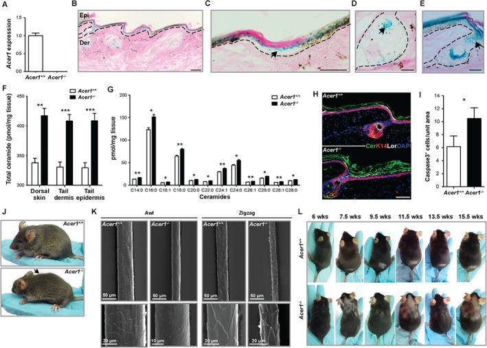
The epidermis is the outermost layer of skin that acts as a barrier to protect the body from the external environment and to control water and heat loss. This barrier function is established through the multistage differentiation of keratinocytes and the presence of bioactive sphingolipids such as ceramides, the levels of which are tightly regulated by a balance of ceramide synthase and ceramidase activities. Here we reveal the essential role of alkaline ceramidase 1 (Acer1) in the skin. Acer1-deficient (Acer1(-/-) ) mice showed elevated levels of ceramide in the skin, aberrant hair shaft cuticle formation and cyclic alopecia. We demonstrate that Acer1 is specifically expressed in differentiated interfollicular epidermis, infundibulum and sebaceous glands and consequently Acer1(-/-) mice have significant alterations in infundibulum and sebaceous gland architecture. Acer1(-/-) skin also shows perturbed hair follicle stem cell compartments. These alterations result in Acer1(-/-) mice showing increased transepidermal water loss and a hypermetabolism phenotype with associated reduction of fat content with age. We conclude that Acer1 is indispensable for mammalian skin homeostasis and whole-body energy homeostasis. © 2016 The Authors. The Journal of Pathology published by John Wiley & Sons Ltd on behalf of Pathological Society of Great Britain and Ireland.

Keywords: ceramidase; energy homeostasis; sebaceous glands; skin homeostasis.


Figures

Figure 1
Figure 1
Mice lacking Acer1 show altered ceramide levels in the skin, hair shaft abnormalities and cyclic alopecia. (A) RT–qPCR analysis of Acer1 expression in tail skin of wild‐type and homozygous mice (n = 4 Acer1 +/+, n = 3 Acer1 −/− females at age 7 weeks). (B–E) LacZ reporter staining shows specific expression of Acer1 in (B) differentiated epidermal layers (Acer1+/−), (C) interfollicular epidermis (arrow; Acer1+/−), (D) sebaceous glands (arrow; Acer1+/−) and (E) infundibulum (arrow; Acer1−/−); Epi, epidermis; Der, dermis. (F) Total ceramide content of dorsal skin, tail epidermis and tail dermis and (G) determination of the individual ceramide species in dorsal skin; data are mean ± standard error (SE) of the mean (n = 5 males, aged 9 weeks) per tissue per genotype; statistical analysis is unpaired t‐test, with adjustment for multiple testing for the individual ceramide species using the Holm–Sidak method with α set to 5%; *p < 0.05, **p < 0.01, ***p < 0.001, ****p < 0.0001. (H) Expression of ceramide (Cer), epidermal basal layer marker keratin 14 (K14) and differentiated layer marker loricrin (Lor) in wild‐type (WT) and Acer1−/− tail epidermis, with nuclear staining (blue) by DAPI (n = 2 males at age 28 weeks/genotype with three technical replicates). (I) Analysis of immunostained skin sections from 28 week‐old Acer1−/− mice shows increased numbers of caspase3+ cells compared to wild‐type mice; data are mean ± SE (n = 2 males/genotype with three technical replicates), statistical analysis was unpaired t‐test, *p = 0.0346. (J) Acer1 −/− mice showed obvious dorsal coat abnormalities (mixed length or long hair, arrow) at age 6 weeks. (K) Representative scanning electron microscopy (SEM) images of different hair types (Awl and Zigzag) from wild‐type and Acer1−/− mice at age 30 weeks (n = 2/genotype). (L) Hair loss associated with hair cycle and age: the small denuded area seen on the lower dorsum of the Acer1 +/+ mouse from 7.5 weeks is the region shaved to perform the hair follicle cycling analysis; this patch has completely regrown in the Acer1 −/− mouse by 7.5 weeks. Scale bars = 100 µm (B–E, H)
Figure 2
Figure 2
The skin of Acer1−/− mice shows hyperproliferation, inflammation and abnormal differentiation. (A) Representative image of H&E staining of tail at different ages (n = 3/age and genotype); open arrows, abnormal SGs in Acer1 −/− skin. (B) Note thickened differentiated layers of the epidermis indicated by black arrows. Immunostaining of skin sections from 27 week‐old Acer1−/− mice shows increased numbers of p63+ cells compared to wild‐type mice; data are shown as mean ± SE (n = 3) and analysed using unpaired t‐test (**p = 0.0017); a representative image is shown; green, p63 staining; blue, nuclear staining by DAPI; Epi, epidermis; Der, dermis. (C) Immunostaining of skin sections from 27 week‐old Acer1−/− mice shows increased numbers of CD45+ cells compared to wild‐type mice; data are shown as mean (n = 3) ± SE and analysed using unpaired t‐test (**p = 0.0216); a representative image is shown; red, CD45 staining; blue, nuclear staining by DAPI; Epi, epidermis; Der, dermis. (D) Expression of epidermal basal layer marker Keratin 14 (K14) and differentiated layer markers filaggrin (Fil) and loricrin (Lor) in Acer1−/− epidermis from different age points (n = 3/age and genotype). (E) TEM images of the epidermal layers: white arrows, abnormal arrangement of cornified layers; black arrows, abnormal keratohyalin (KH) granules in granular layer of Acer1−/− epidermis when compared to wild‐type epidermis; BL, basal layer; SL, suprabasal layer; GL, granular layer; CL, cornified layer; scale bars = 100 µm except in (E) 10 µm
Figure 3
Figure 3
Sebaceous gland and infundibulum expansion in Acer1−/− mice. (A) Representative epidermal whole mount immunostaining of K14 (red) and K15 (green) markers at different ages; blue, DAPI; n = 3/age and genotype). (B) Quantification of total SG area. (C) Representative epidermal whole mount immunostaining of the sebaceous–hair follicle junctional zone marker Lrig1 (green); red, K14; blue, DAPI; n = 3/age and genotype). (D) Quantification of infundibulum width; data are shown as mean ± SE (n = 3) and analysed using unpaired t‐test; ns, not significant; **p = 0.0013, ***p = 0.0010, ****p < 0.0001; scale bars = 100 µm
Figure 4
Figure 4
Sebaceous gland abnormalities in Acer1−/− mice. (A) Representative oil red O staining of epidermal whole mounts at different ages (n = 3/age and genotype). (B, C) TEM images of the SG ultrastructure: note the irregular lipid droplets and nuclear structure in Acer1−/− sebocytes; scale bars = 100 µm, unless stated otherwise
Figure 5
Figure 5
Increased transepidermal water loss and hypermetabolism of Acer1−/− mice. (A) Transepidermal water loss (TEWL) in 19 week‐old male mice (n = 5 Acer1+/+, n = 5 Acer1−/−); data are shown as mean ± SE and analysed using unpaired t‐test; ****p < 0.0001. (B) Energy expenditure (EE; estimated EE for a 30 g mouse, Acer1 −/− versus Acer1 +/+ = 42.7 j/min versus 33.8 j/min; ANCOVA corrected for body weight, p = 5.7 × 10⁻⁹) and (C) food intake [food intake at zero body weight change (dBW): Acer1 −/− versus Acer1 +/+ = 4.5 ± 0.2 g versus 3.7 ± 0.1 g; ANCOVA corrected for change in body weight, p = 0.00116] were measured using indirect calorimetry for 22 h (n = 27 Acer1 +/+, n = 7 Acer1 −/− males). (D) Representative image of H&E‐stained sections of BAT from 18 week‐old male mice (n = 3/genotype); scale bar = 50 µm. (E) Gene expression of Acer1 in adipose tissue depots from 18 week‐old male mice (n = 3/genotype): tail skin RNA from Figure 1A was reused as a positive control for Acer1 detection; nd, not detected. Mean control gene (B2m) C t values for each tissue were: tail skin, 26.9; inguinal WAT, 19.8; epididymal WAT, 19.5; BAT, 22.1

References

    1. Proksch E, Jensen JM, Elias PM. Skin lipids and epidermal differentiation in atopic dermatitis. Clin Dermatol 2003; 21: 134–144. - PubMed
    1. Motta S, Monti M, Sesana S, et al. Ceramide composition of the psoriatic scale. Biochim Biophys Acta 1993; 1182: 147–151. - PubMed
    1. Melnik B, Hollmann J, Plewig G. Decreased stratum corneum ceramides in atopic individuals – a pathobiochemical factor in xerosis? Br J Dermatol 1988; 119: 547–549. - PubMed
    1. Mao C, Xu R, Szulc ZM, et al. Cloning and characterization of a mouse endoplasmic reticulum alkaline ceramidase: an enzyme that preferentially regulates metabolism of very long chain ceramides. J Biol Chem 2003; 278: 31184–31191. - PubMed
    1. Sun W, Xu R, Hu W, et al. Upregulation of the human alkaline ceramidase 1 and acid ceramidase mediates calcium‐induced differentiation of epidermal keratinocytes. J Invest Dermatol 2008; 128: 389–397. - PubMed

Publication types

MeSH terms

LinkOut - more resources