. 2016 Apr 29:6:25264.
doi: 10.1038/srep25264.

Somatic genomic alterations in retinoblastoma beyond RB1 are rare and limited to copy number changes

Affiliations

Somatic genomic alterations in retinoblastoma beyond RB1 are rare and limited to copy number changes

Irsan E Kooi et al. Sci Rep. .

Abstract

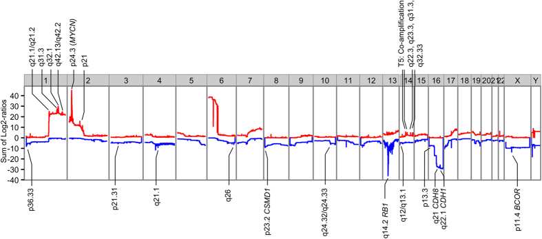
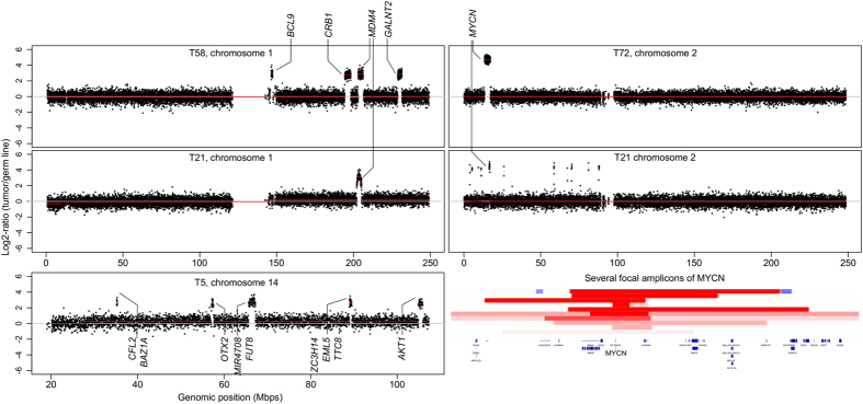
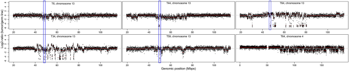
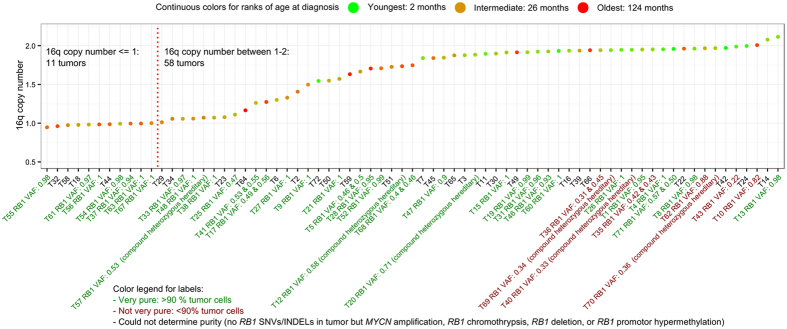
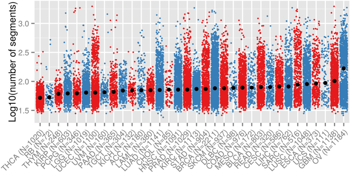
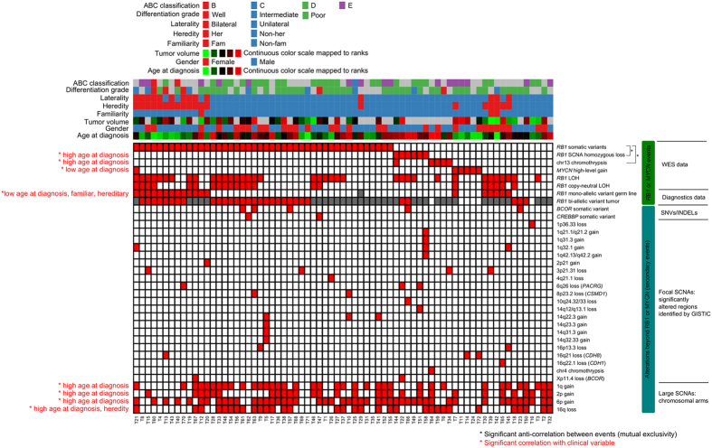
Retinoblastoma is a rare childhood cancer initiated by RB1 mutation or MYCN amplification, while additional alterations may be required for tumor development. However, the view on single nucleotide variants is very limited. To better understand oncogenesis, we determined the genomic landscape of retinoblastoma. We performed exome sequencing of 71 retinoblastomas and matched blood DNA. Next, we determined the presence of single nucleotide variants, copy number alterations and viruses. Aside from RB1, recurrent gene mutations were very rare. Only a limited fraction of tumors showed BCOR (7/71, 10%) or CREBBP alterations (3/71, 4%). No evidence was found for the presence of viruses. Instead, specific somatic copy number alterations were more common, particularly in patients diagnosed at later age. Recurrent alterations of chromosomal arms often involved less than one copy, also in highly pure tumor samples, suggesting within-tumor heterogeneity. Our results show that retinoblastoma is among the least mutated cancers and signify the extreme sensitivity of the childhood retina for RB1 loss. We hypothesize that retinoblastomas arising later in retinal development benefit more from subclonal secondary alterations and therefore, these alterations are more selected for in these tumors. Targeted therapy based on these subclonal events might be insufficient for complete tumor control.


Figures

Figure 1
Figure 1. Significantly altered copy number regions identified by GISTIC.
A karyogram overview of the cumulative copy number changes (Log2-ratios of tumors versus germ line summed over 71 samples, Y-axis) is shown separately for gains (red) and losses (blue). Based on SCNA focality, amplitude and recurrence, GISTIC identified significantly altered regions, which are highlighted by cytoband labels. For regions that contained a single gene only, the gene symbol is given.
Figure 2
Figure 2. Examples of high-level gains.
Segmented (orange lines) somatic copy number estimates (black dots, Log2-ratios, Y-axis) are plotted along genomic coordinates (X-axis). Focal and high-level gains were scarce and were restricted to chromosomes 1 (2/71 tumors), 2 (6/71) and 14 (1/71). Established oncogenes contained in the focal gains are highlighted. Only for chromosome 2, the intersection of amplicons included a single gene only (MYCN). Tumors T58, T21, and T5 showed co-amplification of multiple loci at chromosome 1, 2, and 14 respectively.
Figure 3
Figure 3. Chromothrypsis of chromosome 4 (1/71 tumors) and 13 (5/71 tumors).
Segmented (orange lines) somatic copy number estimates (black dots, Log2-ratios, Y-axis) are plotted along genomic coordinates (X-axis). Chromothrypsis, characterized by clustered chromosomal alterations, was observed for 6 chromosomes in five different tumors. One tumor (T64) showed chromothrypsis at both chromosome 4 and 13. Blue rectangles indicate the RB1 locus.
Figure 4
Figure 4. Amplitudes of 16q loss indicate tumor clonality.
For each tumor (dots) the copy number of 16q is plotted, ordered by increasing copy number. A green-to-red color scale was mapped to age at diagnosis, showing a significant positive association between age at diagnosis and 16q loss amplitude (Kendall’s rank correlation test, p-value 1.2E-06). The tumor labels included VAFs of RB1 variants from which contamination with non-tumor cells can be inferred. Although the majority of tumors were considered very pure (>90%, green labels), 16q loss did rarely reach change of one copy (11 tumors with ploidy <=1), suggesting of within-tumor heterogeneity.
Figure 5
Figure 5. Number of SCNAs across different cancer types.
For each tumor (dots) the number of contiguous DNA segments (so wild type is 22 + X + Y = 24 segments, log10(24) = 0.38) is plotted for our retinoblastoma cohort and all cancer types available at TCGA in alternating colors. Cancer types are ordered by increasing median of SCNAs, showing that the retinoblastoma genome has relatively few SCNAs compared with cancers available in TCGA.
Figure 6
Figure 6. Landscape of somatic alterations in retinoblastoma.
A binary event matrix (red: event occurred, white: event did not occur, grey: data not available) for SNVs/INDELs and SCNA, aligned with color-coded sample information. Columns represent tumor samples and rows represent events. For focal SCNAs containing a single gene only, the gene symbol is given in parentheses. Events that showed significant negative correlation are paired with black lines highlighted on the right of the matrix. Significant correlations between events and clinical variables are indicated by red labels on the left of the matrix. For large SCNAs (1p, 2p, 6p, and 16q), tumors with a mean ploidy more than 2.3 or less than 1.7 were called altered (red boxes).

References

    1. Linabery A. M. & Ross J. A. Trends in childhood cancer incidence in the U.S. (1992–2004). Cancer 112, 416–32 (2008). - PubMed
    1. Parkin D. M., Stiller C. A., Draper G. J. & Bieber C. A. The international incidence of childhood cancer. Int. J. Cancer 42, 511–520 (1988). - PubMed
    1. Rushlow D. E. et al. Characterisation of retinoblastomas without RB1 mutations: genomic, gene expression, and clinical studies. Lancet. Oncol. 14, 327–34 (2013). - PubMed
    1. McEvoy J. et al. RB1 gene inactivation by chromothripsis in human retinoblastoma. Oncotarget 5, 438–50 (2014). - PMC - PubMed
    1. Dimaras H. et al. Loss of RB1 induces non-proliferative retinoma: increasing genomic instability correlates with progression to retinoblastoma. Hum. Mol. Genet. 17, 1363–1372 (2008). - PubMed

Publication types

Substances