Review
. 2017 Jan;45(1):148-163.
doi: 10.1007/s10439-016-1612-8. Epub 2016 Apr 28.

3D Bioprinting for Tissue and Organ Fabrication

Affiliations
Review

3D Bioprinting for Tissue and Organ Fabrication

Yu Shrike Zhang et al. Ann Biomed Eng. 2017 Jan.

Abstract

The field of regenerative medicine has progressed tremendously over the past few decades in its ability to fabricate functional tissue substitutes. Conventional approaches based on scaffolding and microengineering are limited in their capacity of producing tissue constructs with precise biomimetic properties. Three-dimensional (3D) bioprinting technology, on the other hand, promises to bridge the divergence between artificially engineered tissue constructs and native tissues. In a sense, 3D bioprinting offers unprecedented versatility to co-deliver cells and biomaterials with precise control over their compositions, spatial distributions, and architectural accuracy, therefore achieving detailed or even personalized recapitulation of the fine shape, structure, and architecture of target tissues and organs. Here we briefly describe recent progresses of 3D bioprinting technology and associated bioinks suitable for the printing process. We then focus on the applications of this technology in fabrication of biomimetic constructs of several representative tissues and organs, including blood vessel, heart, liver, and cartilage. We finally conclude with future challenges in 3D bioprinting as well as potential solutions for further development.

Keywords: Additive manufacturing; Bioink; Bioprinting; Regenerative medicine; Tissue engineering.


Figures

Figure 1
Figure 1
Approaches for tissue/organ fabrication. (A) Scale of cell numbers encountered in tissue engineering spans at least eight orders of magnitude. The minimum therapeutic threshold for recapitulating solid organ function in humans is estimated at the level of 1-10 billion functioning cells. Reproduced with permission from Ref. , copyright 2014 public library of science. (B) Schematic illustrations of common approaches for tissue engineering. In the scaffold-based approach, cells are seeded into a porous scaffold to populate the matrix and deposit their own ECM. The modular approach, on the other hand, building blocks are utilized to build up large tissue constructs via multiple assembling techniques. Reproduced with permission from Ref. , copyright 2013 Dove Medical Press.
Figure 2
Figure 2
3D Bioprinting with dECM bioinks of different tissue constructs. (A) dECM materials are obtained from various tissues via a multi-step decellularization process that combines physical, chemical and enzymatic treatments. The collected soluble dECM materials are mixed with stem cells and used as bioinks in a layer-by-layer bioprinting approach to fabricate tissue analogues. (B) Native tissues and bioprinted constructs from dECM of the corresponding tissues show similar morphological or histological appearance. Reproduced with permission from Ref. , copyright 2014 Nature Publishing Group.
Figure 3
Figure 3
Bioprinting of vascular structures. (A) Physiology of arteries and veins. Arteries and veins sharing certain features in the multi-layered structures but differ in many other ways. Reproduced with permission from Ref. , copyright 2011 John Wiley & Sons. (B) Construction of macroscale vessels: (i) 3D bioprinted hydrogel; (ii) cross-sectional view; (iii) perspective view of the aorta model. Reproduced with permission from Ref. , copyright 2013 Elsevier. (C) Templated bioprinting based on sacrificial agarose fibers: (i) graphic mode of the agarose template fibers for micromolding; schematic representation of bioprinting of agarose template fibers and subsequent formation of microchannels via template micromolding; (ii) bifurcating bioprinted microchannel network in a GelMA hydrogel; and (iii) confocal image of HUVEC-lined microchannel generated by template micromolding. The inset shows a cross-sectional view of the channel. Scale bars: 250 μm. Reproduced with permission from Ref. , copyright 2014 Royal Society of Chemistry. (D) Schematic views of a heterogeneous bioprinting based on fugitive Pluronics inks: (i) blue filament corresponds to 10T1/2 fibroblast-laden GelMA, red fugitive filament, and green HNDF-laden GelMA ink; (ii) bright-field image of the 3D printed tissue construct, which is overlayed with the green fluorescent channel; (iii) stacked composition of tissue construct. Reproduced with permission from Ref. , copyright 2014 Wiley-VCH. (E) Sacrificial bioprinting based on sugar struts: (i) schematic overview of an bioprinted interconnected, self-supporting carbohydrate-glass lattice; (ii) stacked composition of 10T1/2 uniformly distributed in the fibrin gel and HUVECs in the vascular space; scale bar: 1 mm; (iii) cross-section image of a representative channel; scale bar: 200 μm. Reproduced with permission from Ref. , copyright 2012 Nature Publishing Group.
Figure 4
Figure 4
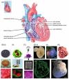
(A) Schematics showing the geographical anatomy of a mature human heart. Reproduced with permission from Ref. , copyright 2011 John Wiley & Sons. (B) Bioprinting of heart valves: (i) heart valve model designed by Solidworks; as-printed valve conduit; Safranin-O staining to stain the glycosaminoglycans red which also stained the Me-HA within the hydrogel red. Reproduced with permission from Ref. , copyright 2014 Elsevier. (ii) Heterogeneous aortic valve e-printing software sliced the geometries into layers and generated extrusion paths for each layer along with viable HAVIC-seeded valve scaffolds containing cells across the entire surface of the conduits. Reproduced with permission from Ref. , copyright 2013 Wiley-VCH. (C) Cardiac cells bioprinted in decelluarized cardiac extracellular matrix. Scale bars: 5 mm and 400 μm. Reproduced with permission from Ref. , copyright 2014 Nature Publishing Group. (D) Bioprinting of the whole-heart structure: (i) A darkfield image of an explanted embryonic chick heart. (ii) A confocal fluorescence micrograph of the heart stained for fibronectin (green), nuclei (blue), and F-actin (red). (iii) The 3D CAD model of the heart with complex internal architecture based on the confocal data. (iv) A cross section of the 3D bioprinted heart showing recreation of the internal trabecular structure from the CAD model. (v) A dark-field image of the 3D printed heart with internal structure visible through the translucent heart wall. Scale bars: 1 mm in (i) and (ii) and 1 cm in (iii) and (iv). Reproduced with permission from Ref. , copyright 2015 American Association for the Advancement of Science.
Figure 5
Figure 5
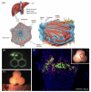
Bioprinting of liver tissues. (A) Layout of typical structural units of the hepatic lobule. In cross-sectional views, the microstructures appear as a hexagonal lattice, with the hepatic artery, bile duct, and portal vein triads placed at the hexagon vertices. Reproduced with permission from Ref. , copyright 2011 John Wiley & Sons. (B-D) Bioprinted liver tissue constructs with similar arrangement of the hepatic lobules to native liver tissues and tissue-like cellular density and tight intercellular junctions, using human primary hepatocytes, endothelial cells, and hepatic stellate cells. (B, C) Photographs showing the liver organoids immediately after bioprinting. (D) Fluorescence micrograph of the planar cross-section after tissue maturation, highlighting the compartmentalization of the non-parenchymal cells relative to the hepatocytes. The hepatic stellate cells and endothelial cells were pre-labeled in green and red, respectively, while the nuclei of all cells were stained in blue. Adapted with permission from Ref. , copyright 2015 OrganovoTM.

References

    1. Langer R, Vacanti JP. Tissue engineering. Science. 1993;260:920–926. - PubMed
    1. Langer R, Vacanti JP, Vacanti CA, Atala A, Freed LE, Vunjak-Novakovic G. Tissue engineering: Biomedical applications. Tissue Eng. 1995;1:151–161. - PubMed
    1. Langer R. Tissue engineering: Status and challenges. e-Biomed: J Regen Med. 2000;1:5–6.
    1. Khademhosseini A, Vacanti JP, Langer R. Progress in tissue engineering. Sci Am. 2009;300:64–71. - PubMed
    1. Rice JJ, Martino MM, De Laporte L, Tortelli F, Briquez PS, Hubbell JA. Engineering the regenerative microenvironment with biomaterials. Adv Healthc Mater. 2013;2:57–71. - PubMed

Publication types