Review
. 2016 Jun;57(6):827-33.
doi: 10.2967/jnumed.115.172023. Epub 2016 Apr 28.

Molecular Imaging of Ovarian Cancer

Affiliations
Review

Molecular Imaging of Ovarian Cancer

Sai Kiran Sharma et al. J Nucl Med. 2016 Jun.

Abstract

Ovarian cancer is the most lethal gynecologic malignancy and the fifth leading cause of cancer-related death in women. Over the past decade, medical imaging has played an increasingly valuable role in the diagnosis, staging, and treatment planning of the disease. In this "Focus on Molecular Imaging" review, we seek to provide a brief yet informative survey of the current state of the molecular imaging of ovarian cancer. The article is divided into sections according to modality, covering recent advances in the MR, PET, SPECT, ultrasound, and optical imaging of ovarian cancer. Although primary emphasis is given to clinical studies, preclinical investigations that are particularly innovative and promising are discussed as well. Ultimately, we are hopeful that the combination of technologic innovations, novel imaging probes, and further integration of imaging into clinical protocols will lead to significant improvements in the survival rate for ovarian cancer.

Keywords: MRI; PET; SPECT; molecular imaging; ovarian cancer; ultrasound.


Conflict of interest statement

No other potential conflict of interest relevant to this article was reported.

Figures

FIGURE 1
FIGURE 1
Multiparametric pretreatment MR images (3.0 T) of primary ovarian tumor (red arrows), omental cake (blue arrows), and peritoneal implant (green arrows) in 58-y-old woman with advanced ovarian cancer: T2-weighted image (A); diffusion-weighted image (b = 5 500 s/mm2) (B); fused T2-weighted and diffusion-weighted image (C); T2-weighted image showing region of interest for MRS (red) from primary tumor (D); spectral fit (red) overlaid on raw MRS data (gray), illustrating strong choline (Cho) signal (E). (Adapted and reprinted with permission of (12).)
FIGURE 2
FIGURE 2
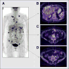
18F-FDG PET/CT images of recurrent ovarian cancer 10 mo after treatment with radical surgery and adjuvant chemotherapy: maximum-intensity projection (A) and transaxial images delineating uptake of radiotracer in liver (B), peritoneum (C), and locoregional lymph nodes (D). (Adapted and reprinted with permission of (16).)
FIGURE 3
FIGURE 3
Identification of metastatic ovarian cancer deposits via intraoperative and ex vivo fluorescence imaging. (A and B) Comparative images of tumors within abdominal cavity visualized under white light (A) vs. fluorescence emissions from folate receptor–targeted probe (B). (C) Statistical analysis revealing benefit of intraoperative fluorescence imaging. (D) Identification of two metastatic lesions in greater omentum using indocyanine green dye for intraoperative near-infrared (NIR) fluorescence imaging. (E) Ex vivo visualization of resected metastatic lesions. (Adapted and reprinted with permission of (31,33).)
FIGURE 4
FIGURE 4
Preclinical contrast-enhanced ultrasound imaging of angiogenesis. Microbubbles functionalized with CD276-targeting antibody (MBCD276) effectively targeted subcutaneous xenografts comprising 2008 human endometrioid ovarian cancer cells mixed with CD276-expressing MS1 mouse endothelial cells. Specificity of targeting was demonstrated via comparison with isotype antibody-functionalized microbubbles (MBISO) as well as blocking experiment in which excess of CD276 antibody was administered. (Adapted and reprinted with permission of (42).)
FIGURE 5
FIGURE 5
PET/MRI, an emerging hybrid imaging technique: axial (A) and coronal (B) T2-weighted MRI scans of ovarian cancer patient, in which tumor lesions were seen adjacent to liver (long solid arrow), in segment IV of liver (dotted arrow), in porta hepatis (short solid arrow), and in peritoneum (arrowheads); axial (C) and coronal (D) PET/MRI scans wherein 18F-FDG PET not only demonstrated excellent correlation with lesions identified previously by MRI alone but also highlighted new lesions (arrows); (E) whole-body maximum-intensity projection from 18F-FDG PET showing multiple lesions in chest (arrow) and abdomen. (Adapted and reprinted with permission of (43).)

References

    1. Siegel RL, Miller KD, Jemal A. Cancer statistics, 2015. CA Cancer J Clin. 2015;65:5–29. - PubMed
    1. Gupta D, Lis CG. Role of CA125 in predicting ovarian cancer survival: a review of the epidemiological literature. J Ovarian Res. 2009;2:13. - PMC - PubMed
    1. Glaser G, Torres M, Kim BH, et al. The use of CT findings to predict extent of tumor at primary surgery for ovarian cancer. Gynecol Oncol. 2013;130:280–283. - PMC - PubMed
    1. Iyer VR, Lee SI. MRI, CT, and PET/CT for ovarian cancer detection and adnexal lesion characterization. AJR. 2010;194:311–321. - PubMed
    1. Burger IA, Goldman DA, Vargas HA, et al. Incorporation of postoperative CT data into clinical models to predict 5-year overall and recurrence free survival after primary cytoreductive surgery for advanced ovarian cancer. Gynecol Oncol. 2015;138:554–559. - PMC - PubMed