

Protease-activated receptors in hemostasis
- PMID: 27127302
- PMCID: PMC4946198
- DOI: 10.1182/blood-2015-11-636472
Protease-activated receptors in hemostasis
Abstract
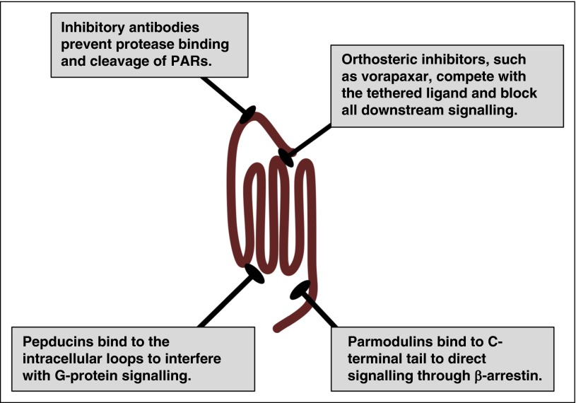
Protease signaling in cells elicits multiple physiologically important responses via protease-activated receptors (PARs). There are 4 members of this family of G-protein-coupled receptors (PAR1-4). PARs are activated by proteolysis of the N terminus to reveal a tethered ligand. The rate-limiting step of PAR signaling is determined by the efficiency of proteolysis of the N terminus, which is regulated by allosteric binding sites, cofactors, membrane localization, and receptor dimerization. This ultimately controls the initiation of PAR signaling. In addition, these factors also control the cellular response by directing signaling toward G-protein or β-arrestin pathways. PAR1 signaling on endothelial cells is controlled by the activating protease and heterodimerization with PAR2 or PAR3. As a consequence, the genetic and epigenetic control of PARs and their cofactors in physiologic and pathophysiologic conditions have the potential to influence cellular behavior. Recent studies have uncovered polymorphisms that result in PAR4 sequence variants with altered reactivity that interact to influence platelet response. This further demonstrates how interactions within the plasma membrane can control the physiological output. Understanding the structural rearrangement following PAR activation and how PARs are allosterically controlled within the plasma membrane will determine how best to target this family of receptors therapeutically. The purpose of this article is to review how signaling from PARs is influenced by alternative cleavage sites and the physical interactions within the membrane. Going forward, it will be important to relate the altered signaling to the molecular arrangement of PARs in the cell membrane and to determine how these may be influenced genetically.
© 2016 by The American Society of Hematology.
Figures
References
-
- Vu TK, Hung DT, Wheaton VI, Coughlin SR. Molecular cloning of a functional thrombin receptor reveals a novel proteolytic mechanism of receptor activation. Cell. 1991;64(6):1057–1068. - PubMed
-
- O’Brien PJ, Molino M, Kahn M, Brass LF. Protease activated receptors: theme and variations. Oncogene. 2001;20(13):1570–1581. - PubMed
-
- Ishihara H, Connolly AJ, Zeng D, et al. Protease-activated receptor 3 is a second thrombin receptor in humans. Nature. 1997;386(6624):502–506. - PubMed
-
- Kahn ML, Zheng YW, Huang W, et al. A dual thrombin receptor system for platelet activation. Nature. 1998;394(6694):690–694. - PubMed
Publication types
MeSH terms
Substances
Grants and funding
LinkOut - more resources
Full Text Sources
Other Literature Sources
Research Materials
Miscellaneous
