

An Undescribed Monteggia Type 3 Equivalent Lesion: Lateral Dislocation of Radial Head with Both-Bone Forearm Fracture
- PMID: 27127669
- PMCID: PMC4834152
- DOI: 10.1155/2016/8598139
An Undescribed Monteggia Type 3 Equivalent Lesion: Lateral Dislocation of Radial Head with Both-Bone Forearm Fracture
Abstract
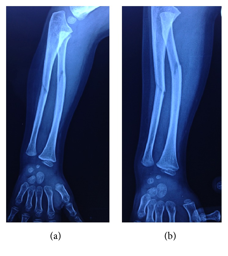
Monteggia fractures are accepted as hard-to-recognize and easy-to-handle fractures. Adequate radiographic investigations and clinical examinations are necessities. This case holds unique features involving diagnosis and treatment. In this case, the radial head was dislocated laterally while both bones were fractured in the proximal diaphysis, being the first to be mentioned in the literature. Closed reduction of the ulna is the preferred method of handling and almost always results in reduction of the radial head. Literature obligates ulnar reduction as a preliminary to reduce and stabilize the radial head. Closed reduction reduced the ulna but the radial head was not reduced. Hence an intramedullary K-wire was used to reduce the radial head and a long arm cast was used to stabilize the reduction. The operation was successful and follow-up showed no complications.
Figures


References
-
- Herring J. A., Ho C. Upper extremity injuries. In: Herring J. A., editor. Tachdjian's Pediatric Orthopaedics. Philadelphia, Pa, USA: Elsevier Saunders; 2014. pp. 1326–1332.
-
- Dolan M., Waters P. Fractures and dislocations of the forearm, wrist, and hand. In: Green N. E., Swiontkowski M. F., editors. Skeletal Trauma in Children. Philadelphia, Pa, USA: Elsevier Saunders; 2009. pp. 182–184.
LinkOut - more resources
Full Text Sources
Other Literature Sources
Miscellaneous
