

Semiautomatic Segmentation of Rim Area Focal Hyperautofluorescence Predicts Progression of Geographic Atrophy Due to Dry Age-Related Macular Degeneration
- PMID: 27127926
- PMCID: PMC5221410
- DOI: 10.1167/iovs.15-19008
Semiautomatic Segmentation of Rim Area Focal Hyperautofluorescence Predicts Progression of Geographic Atrophy Due to Dry Age-Related Macular Degeneration
Abstract
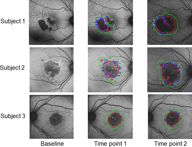
Purpose: To develop image analysis software usable by nonexpert graders to segment geographic atrophy (GA) from dry AMD and to quantify rim area focal hyperautofluorescence (RAFH) surrounding GA on fundus autofluorescence (FAF) images. To compare the GA progression predictions based on RAFH with those of a validated qualitative classification system.
Methods: Retrospective analysis of serial FAF images from 49 eyes of 30 subjects with GA was performed using MATLAB-based software (MathWorks, Natick, MA, USA). Correlation between RAFH and progression of GA was analyzed using Spearman correlation. Comparisons of lesion growth rate between RAFH tertiles used generalized estimating equations and Kruskal-Wallis testing. Interobserver variability in lesion size, growth rate and RAFH were compared between two expert and one nonexpert grader using Bland-Altman statistics.
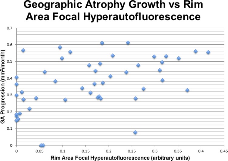
Results: Rim area focal hyperautofluorescence was positively correlated with GA progression rate (ρ = 0.49, P < 0.001). Subjects in the middle or highest RAFH tertile were at greater risk of progression (P = 0.005 and P = 0.001, respectively). Mean difference in RAFH was 0.012 between expert and -0.005 to 0.017 between expert and nonexperts. Mean difference in lesion size (mm2) was 0.11 between expert and -0.29 to 0.41 between expert and nonexperts. Mean difference in lesion growth rate (mm2/mo) was 0.0098 between expert and -0.027 to 0.037 between expert and nonexperts. Risk stratification based on RAFH tertile was 96% identical across all graders.
Conclusions: Our semiautomated image analysis software facilitates stratification of progression risk based on RAFH and enabled a nonexpert grader with minimal training to obtain results comparable to expert graders. Predictions based on RAFH were similar to those of a validated qualitative classification system.
Figures
Comment in
-
Understanding RPE Lipofuscin.Invest Ophthalmol Vis Sci. 2016 Dec 1;57(15):6766. doi: 10.1167/iovs.16-21081. Invest Ophthalmol Vis Sci. 2016. PMID: 27978557 No abstract available.
Similar articles
-
Association of Hyperautofluorescence Signals with Geographic Atrophy Progression in the METformin for the MINimization of Geographic Atrophy Progression Trial.Ophthalmol Sci. 2024 Sep 12;5(1):100620. doi: 10.1016/j.xops.2024.100620. eCollection 2025 Jan-Feb. Ophthalmol Sci. 2024. PMID: 39584185 Free PMC article.
-
Segmentation of the geographic atrophy in spectral-domain optical coherence tomography and fundus autofluorescence images.Invest Ophthalmol Vis Sci. 2013 Dec 30;54(13):8375-83. doi: 10.1167/iovs.13-12552. Invest Ophthalmol Vis Sci. 2013. PMID: 24265015
-
Semiautomated image processing method for identification and quantification of geographic atrophy in age-related macular degeneration.Invest Ophthalmol Vis Sci. 2011 Sep 29;52(10):7640-6. doi: 10.1167/iovs.11-7457. Invest Ophthalmol Vis Sci. 2011. PMID: 21873669
-
Fundus autofluorescence imaging in age-related macular degeneration and geographic atrophy.Adv Exp Med Biol. 2010;664:395-402. doi: 10.1007/978-1-4419-1399-9_45. Adv Exp Med Biol. 2010. PMID: 20238040 Review.
-
RECLASSIFICATION OF FUNDUS AUTOFLUORESCENCE PATTERNS SURROUNDING GEOGRAPHIC ATROPHY BASED ON PROGRESSION RATE: A Systematic Review and Meta-Analysis.Retina. 2019 Oct;39(10):1829-1839. doi: 10.1097/IAE.0000000000002480. Retina. 2019. PMID: 30829988
Cited by
-
Novel Image-Based Analysis for Reduction of Clinician-Dependent Variability in Measurement of the Corneal Ulcer Size.Cornea. 2018 Mar;37(3):331-339. doi: 10.1097/ICO.0000000000001488. Cornea. 2018. PMID: 29256985 Free PMC article.
-
Geographic atrophy: Understanding the relationship between structure and function.Asia Pac J Ophthalmol (Phila). 2025 May-Jun;14(3):100207. doi: 10.1016/j.apjo.2025.100207. Epub 2025 May 19. Asia Pac J Ophthalmol (Phila). 2025. PMID: 40398512 Review.
-
Visible light OCT-based quantitative imaging of lipofuscin in the retinal pigment epithelium with standard reference targets.Biomed Opt Express. 2018 Jul 23;9(8):3768-3782. doi: 10.1364/BOE.9.003768. eCollection 2018 Aug 1. Biomed Opt Express. 2018. PMID: 30338154 Free PMC article.
-
Identifying retinal pigment epithelium cells in adaptive optics-optical coherence tomography images with partial annotations and superhuman accuracy.Biomed Opt Express. 2024 Nov 21;15(12):6922-6939. doi: 10.1364/BOE.538473. eCollection 2024 Dec 1. Biomed Opt Express. 2024. PMID: 39679394 Free PMC article.
-
Fundus Autofluorescence Variation in Geographic Atrophy of Age-Related Macular Degeneration: A Clinicopathologic Correlation.Invest Ophthalmol Vis Sci. 2025 Jan 2;66(1):49. doi: 10.1167/iovs.66.1.49. Invest Ophthalmol Vis Sci. 2025. PMID: 39836402 Free PMC article.
References
-
- Klein R. Prevalence of age-related macular degeneration in the US population. Arch Ophthalmol. 2011; 129: 75. - PubMed
-
- Blair CJ. Geographic atrophy of the retinal pigment epithelium. A manifestation of senile macular degeneration. Arch Ophthalmol. 1975; 93: 19–25. - PubMed
-
- Giocanti-Auregan A,, Tadayoni R,, Fajnkuchen F,, Dourmad P,, Magazzeni S,, Cohen SY. Predictive value of outer retina en face OCT imaging for geographic atrophy progression. Invest Ophthalmol Vis Sci. 2015; 56: 8325–8330. - PubMed
-
- Nunes RP,, Gregori G,, Yehoshua Z,, et al. Predicting the progression of geographic atrophy in age-related macular degeneration with SD-OCT en face imaging of the outer retina. Ophthalmic Surg Lasers Imaging Retina. 44: 344–359. - PubMed
-
- Folgar FA,, Yuan EL,, Sevilla MB,, et al. Drusen volume and retinal pigment epithelium abnormal thinning volume predict 2-year progression of age-related macular degeneration. Ophthalmology. 2016; 123: 39–50, e1. - PubMed
MeSH terms
Grants and funding
LinkOut - more resources
Full Text Sources
Other Literature Sources
