

Antioxidant Treatment Limits Neuroinflammation in Experimental Glaucoma
- PMID: 27127934
- PMCID: PMC4855827
- DOI: 10.1167/iovs.16-19153
Antioxidant Treatment Limits Neuroinflammation in Experimental Glaucoma
Abstract
Purpose: Besides primary neurotoxicity, oxidative stress may compromise the glial immune regulation and shift the immune homeostasis toward neurodegenerative inflammation in glaucoma. We tested this hypothesis through the analysis of neuroinflammatory and neurodegenerative outcomes in mouse glaucoma using two experimental paradigms of decreased or increased oxidative stress.
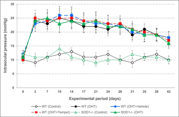
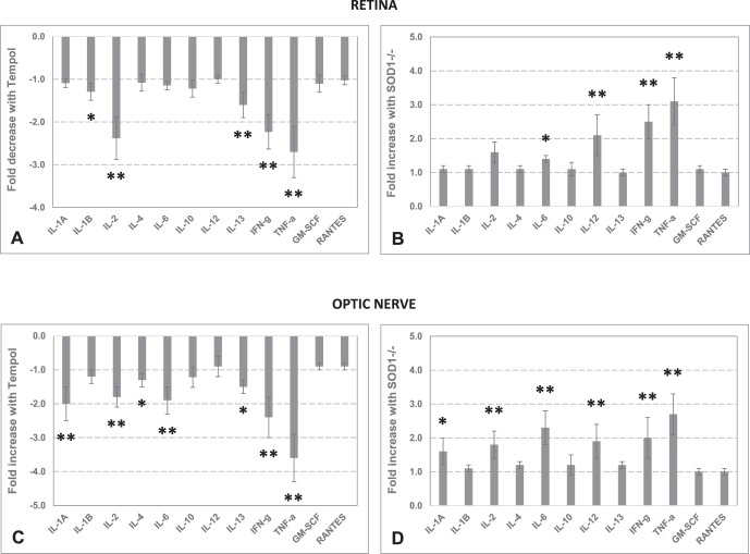
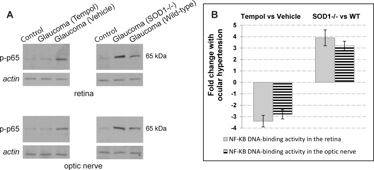
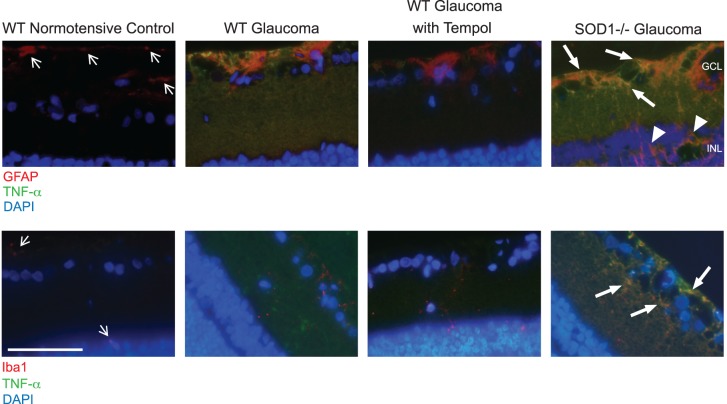
Methods: The first experimental paradigm tested the effects of Tempol, a multifunctional antioxidant, given through osmotic mini-pumps for drug delivery by constant infusion. Following a 6-week treatment period after microbead/viscoelastic injection-induced ocular hypertension, retina and optic nerve samples were analyzed for markers of oxidative stress and cytokine profiles using specific bioassays. We also analyzed a redox-sensitive transcriptional regulator of neuroinflammation, namely NF-κB. The second paradigm included a similar analysis of the effects of overloaded oxidative stress on retina and optic nerve inflammation in mice knockout for a major antioxidant enzyme (SOD1(-/-)).
Results: Increased antioxidant capacity and decreased protein carbonyls and HNE adducts with Tempol treatment verified the drug delivery and biological function. Among a range of cytokines measured, proinflammatory cytokines, including IL-1, IL-2, IFN-γ, and TNF-α, exhibited more than 2-fold decreased titers in Tempol-treated ocular hypertensive eyes. Antioxidant treatment also resulted in a prominent decrease in NF-κB activation in the ocular hypertensive retina and optic nerve. Although pharmacological treatment limiting the oxidative stress resulted in decreased neuroinflammation, ocular hypertension-induced neuroinflammatory responses were increased in SOD1(-/-) mice with defective antioxidant response.
Conclusions: These findings support the oxidative stress-related mechanisms of neuroinflammation and the potential of antioxidant treatment as an immunomodulation strategy for neuroprotection in glaucoma.
Figures


Similar articles
-
cFLIP in the molecular regulation of astroglia-driven neuroinflammation in experimental glaucoma.J Neuroinflammation. 2024 Jun 1;21(1):145. doi: 10.1186/s12974-024-03141-4. J Neuroinflammation. 2024. PMID: 38824526 Free PMC article.
-
Transgenic inhibition of astroglial NF-κB restrains the neuroinflammatory and neurodegenerative outcomes of experimental mouse glaucoma.J Neuroinflammation. 2020 Aug 28;17(1):252. doi: 10.1186/s12974-020-01930-1. J Neuroinflammation. 2020. PMID: 32859212 Free PMC article.
-
Fisetin rescues retinal functions by suppressing inflammatory response in a DBA/2J mouse model of glaucoma.Doc Ophthalmol. 2019 Apr;138(2):125-135. doi: 10.1007/s10633-019-09676-9. Epub 2019 Feb 11. Doc Ophthalmol. 2019. PMID: 30756213
-
Neuroinflammation in Glaucoma and Optic Nerve Damage.Prog Mol Biol Transl Sci. 2015;134:343-63. doi: 10.1016/bs.pmbts.2015.06.010. Epub 2015 Jul 10. Prog Mol Biol Transl Sci. 2015. PMID: 26310164 Review.
-
Clinical potential of lomerizine, a Ca2+ channel blocker as an anti-glaucoma drug: effects on ocular circulation and retinal neuronal damage.Cardiovasc Drug Rev. 2004 Fall;22(3):199-214. doi: 10.1111/j.1527-3466.2004.tb00141.x. Cardiovasc Drug Rev. 2004. PMID: 15492768 Review.
Cited by
-
Transcorneal Electrical Stimulation Inhibits Retinal Microglial Activation and Enhances Retinal Ganglion Cell Survival After Acute Ocular Hypertensive Injury.Transl Vis Sci Technol. 2018 May 29;7(3):7. doi: 10.1167/tvst.7.3.7. eCollection 2018 May. Transl Vis Sci Technol. 2018. PMID: 29862139 Free PMC article.
-
Challenging glaucoma with emerging therapies: an overview of advancements against the silent thief of sight.Front Med (Lausanne). 2025 Mar 26;12:1527319. doi: 10.3389/fmed.2025.1527319. eCollection 2025. Front Med (Lausanne). 2025. PMID: 40206485 Free PMC article. Review.
-
Crosstalk Between Dysfunctional Mitochondria and Inflammation in Glaucomatous Neurodegeneration.Front Pharmacol. 2021 Jul 21;12:699623. doi: 10.3389/fphar.2021.699623. eCollection 2021. Front Pharmacol. 2021. PMID: 34366851 Free PMC article. Review.
-
Chronic Antioxidant Capacity Loss in Anterior Chamber Environment After Iridectomy.Transl Vis Sci Technol. 2023 May 1;12(5):4. doi: 10.1167/tvst.12.5.4. Transl Vis Sci Technol. 2023. PMID: 37126333 Free PMC article.
-
Candidate SNP Markers Significantly Altering the Affinity of the TATA-Binding Protein for the Promoters of Human Genes Associated with Primary Open-Angle Glaucoma.Int J Mol Sci. 2024 Nov 28;25(23):12802. doi: 10.3390/ijms252312802. Int J Mol Sci. 2024. PMID: 39684516 Free PMC article.
References
-
- Quigley HA. Glaucoma: macrocosm to microcosm the Friedenwald lecture. Invest Ophthalmol Vis Sci. 2005; 46: 2662–2670. - PubMed
-
- Quigley HA,, Vitale S. Models of open-angle glaucoma prevalence and incidence in the United States. Invest Ophthalmol Vis Sci. 1997; 38: 83–91. - PubMed
-
- Libby RT,, Gould DB,, Anderson MG,, John SW. Complex genetics of glaucoma susceptibility. Annu Rev Genomics Hum Genet. 2005; 6: 15–44. - PubMed
Publication types
MeSH terms
Substances
Grants and funding
LinkOut - more resources
Full Text Sources
Other Literature Sources
Medical
Molecular Biology Databases
Miscellaneous
