

Assessment of Glioma Response to Radiotherapy Using Multiple MRI Biomarkers with Manual and Semiautomated Segmentation Algorithms
- PMID: 27128445
- PMCID: PMC5083211
- DOI: 10.1111/jon.12354
Assessment of Glioma Response to Radiotherapy Using Multiple MRI Biomarkers with Manual and Semiautomated Segmentation Algorithms
Abstract
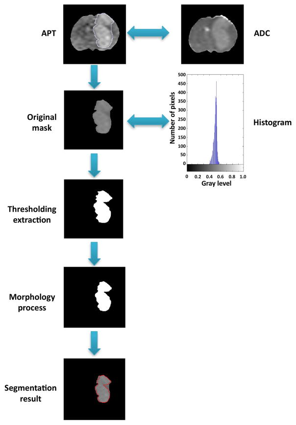
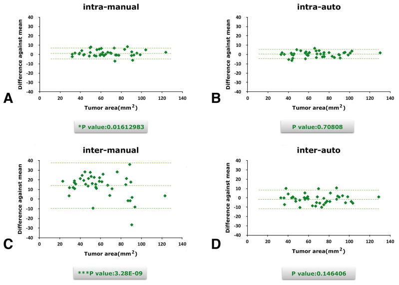
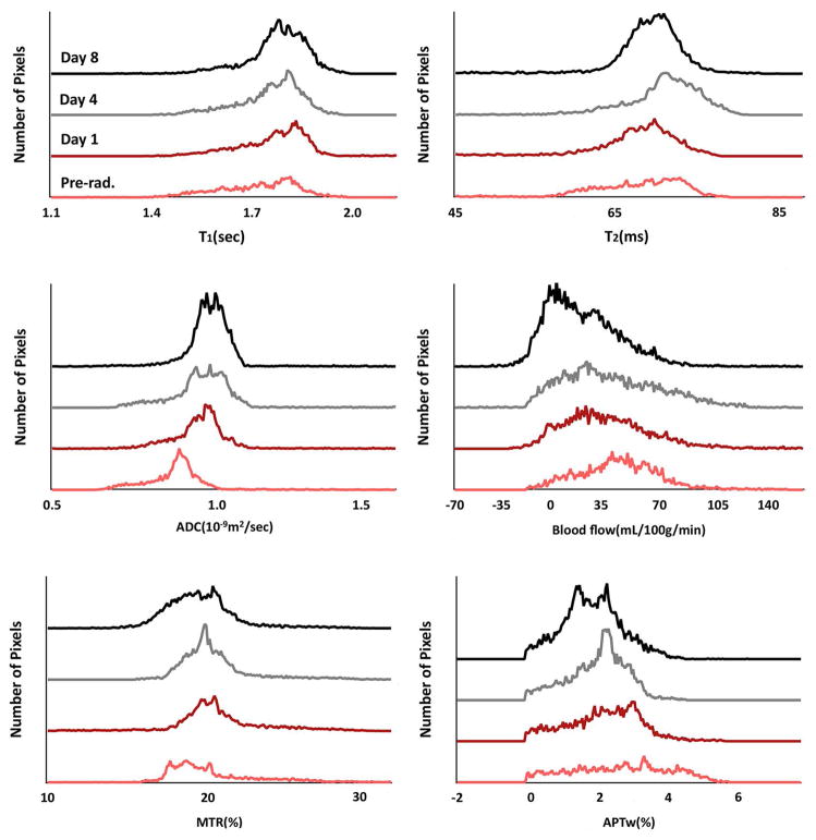
Background and purpose: Multimodality magnetic resonance imaging (MRI) can provide complementary information in the assessment of brain tumors. We aimed to segment tumor in amide proton transfer-weighted (APTw) images and to investigate multiparametric MRI biomarkers for the assessment of glioma response to radiotherapy. For tumor extraction, we evaluated a semiautomated segmentation method based on region of interest (ROI) results by comparing it with the manual segmentation method.
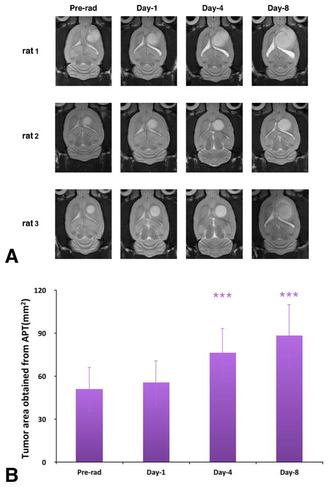
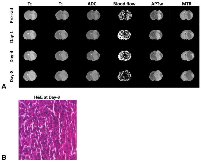
Methods: Thirteen nude rats injected with U87 tumor cells were irradiated by an 8-Gy radiation dose. All MRI scans were performed on a 4.7-T animal scanner preradiation, and at day 1, day 4, and day 8 postradiation. Two experts performed manual and semiautomated methods to extract tumor ROIs on APTw images. Multimodality MRI signals of the tumors, including structural (T2 and T1 ), functional (apparent diffusion coefficient and blood flow), and molecular (APTw and magnetization transfer ratio or MTR), were calculated and compared quantitatively.
Results: The semiautomated method provided more reliable tumor extraction results on APTw images than the manual segmentation, in less time. A considerable increase in the ADC intensities of the tumor was observed during the postradiation. A steady decrease in the blood flow values and in the APTw signal intensities were found after radiotherapy.
Conclusions: The semiautomated method of tumor extraction showed greater efficiency and stability than the manual method. Apparent diffusion coefficient, blood flow, and APTw are all useful biomarkers in assessing glioma response to radiotherapy.
Keywords: APT imaging; Brain tumor; image segmentation; multiparametric MRI; response assessment.
Copyright © 2016 by the American Society of Neuroimaging.
Figures
Similar articles
-
Quantitative multiparametric MRI assessment of glioma response to radiotherapy in a rat model.Neuro Oncol. 2014 Jun;16(6):856-67. doi: 10.1093/neuonc/not245. Epub 2013 Dec 22. Neuro Oncol. 2014. PMID: 24366911 Free PMC article.
-
Quantitative correlational study of microbubble-enhanced ultrasound imaging and magnetic resonance imaging of glioma and early response to radiotherapy in a rat model.Med Phys. 2015 Aug;42(8):4762-72. doi: 10.1118/1.4926550. Med Phys. 2015. PMID: 26233204 Free PMC article.
-
Repeatability of amide proton transfer-weighted signals in the brain according to clinical condition and anatomical location.Eur Radiol. 2020 Jan;30(1):346-356. doi: 10.1007/s00330-019-06285-7. Epub 2019 Jul 23. Eur Radiol. 2020. PMID: 31338651
-
Amide proton transfer-weighted MRI in distinguishing high- and low-grade gliomas: a systematic review and meta-analysis.Neuroradiology. 2019 May;61(5):525-534. doi: 10.1007/s00234-018-02152-2. Epub 2019 Jan 21. Neuroradiology. 2019. PMID: 30666352
-
Multi-parametric MR Imaging Biomarkers Associated to Clinical Outcomes in Gliomas: A Systematic Review.Curr Med Imaging Rev. 2019;15(10):933-947. doi: 10.2174/1573405615666190109100503. Curr Med Imaging Rev. 2019. PMID: 32008521
Cited by
-
Implementing diffusion-weighted MRI for body imaging in prospective multicentre trials: current considerations and future perspectives.Eur Radiol. 2018 Mar;28(3):1118-1131. doi: 10.1007/s00330-017-4972-z. Epub 2017 Sep 27. Eur Radiol. 2018. PMID: 28956113 Free PMC article. Review.
-
Magnetic resonance imaging-guided radiation therapy using animal models of glioblastoma.Br J Radiol. 2019 Mar;92(1095):20180713. doi: 10.1259/bjr.20180713. Epub 2019 Jan 17. Br J Radiol. 2019. PMID: 30563357 Free PMC article. Review.
-
Rapid and quantitative chemical exchange saturation transfer (CEST) imaging with magnetic resonance fingerprinting (MRF).Magn Reson Med. 2018 Dec;80(6):2449-2463. doi: 10.1002/mrm.27221. Epub 2018 May 13. Magn Reson Med. 2018. PMID: 29756286 Free PMC article.
-
Evaluation of Temozolomide Treatment for Glioblastoma Using Amide Proton Transfer Imaging and Diffusion MRI.Cancers (Basel). 2022 Apr 10;14(8):1907. doi: 10.3390/cancers14081907. Cancers (Basel). 2022. PMID: 35454814 Free PMC article.
-
Current Applications and Future Development of Magnetic Resonance Fingerprinting in Diagnosis, Characterization, and Response Monitoring in Cancer.Cancers (Basel). 2021 Sep 22;13(19):4742. doi: 10.3390/cancers13194742. Cancers (Basel). 2021. PMID: 34638229 Free PMC article. Review.
References
-
- Legler JM, Ries LA, Smith MA, et al. Cancer surveillance series [corrected]: brain and other central nervous system cancers: recent trends in incidence and mortality. J Natl Cancer Inst. 1999;91:1382–90. - PubMed
-
- Wen PY, Kesari S. Malignant gliomas in adults. N Engl J Med. 2008;359:492–507. - PubMed
-
- Walker MD, Strike TA, Sheline GE. An analysis of dose-effect relationship in the radiotherapy of malignant gliomas. Int J Radiat Oncol Biol Phys. 1979;5:1725–31. - PubMed
-
- Macdonald DR, Cascino TL, Schold SC, Jr, Cairncross JG. Response criteria for phase II studies of supratentorial malignant glioma. J Clin Oncol. 1990;8:1277–80. - PubMed
-
- Wen PY, Macdonald DR, Reardon DA, et al. Updated response assessment criteria for high-grade gliomas: response assessment in neuro-oncology working group. J Clin Oncol. 2010;28:1963–72. - PubMed
MeSH terms
Substances
Grants and funding
LinkOut - more resources
Full Text Sources
Other Literature Sources
Medical
