. 2016 Apr 29;12(4):e1005600.
doi: 10.1371/journal.ppat.1005600. eCollection 2016 Apr.

Distinct Roles of Type I and Type III Interferons in Intestinal Immunity to Homologous and Heterologous Rotavirus Infections

Affiliations

Distinct Roles of Type I and Type III Interferons in Intestinal Immunity to Homologous and Heterologous Rotavirus Infections

Jian-Da Lin et al. PLoS Pathog. .

Erratum in

Abstract

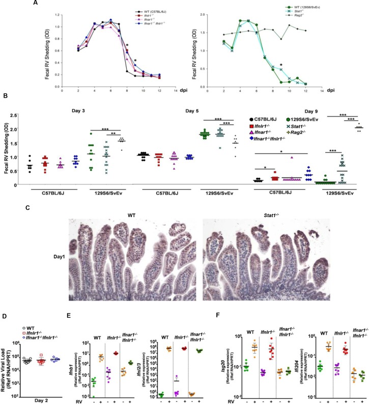
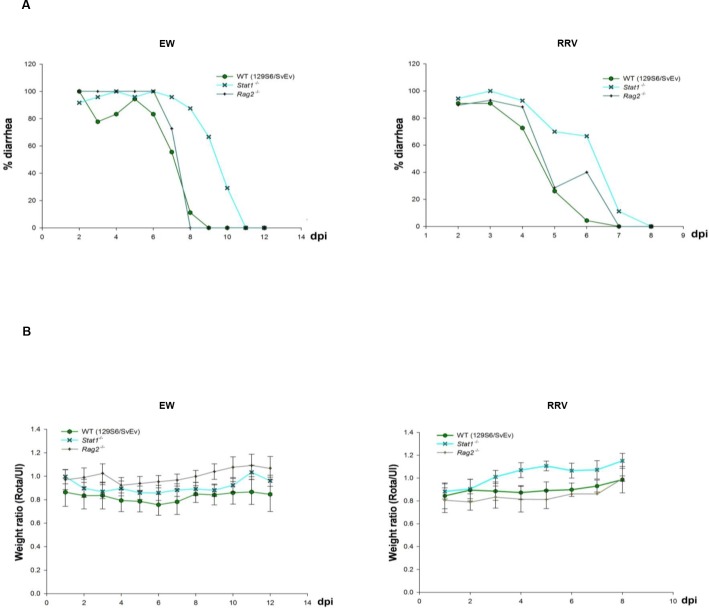
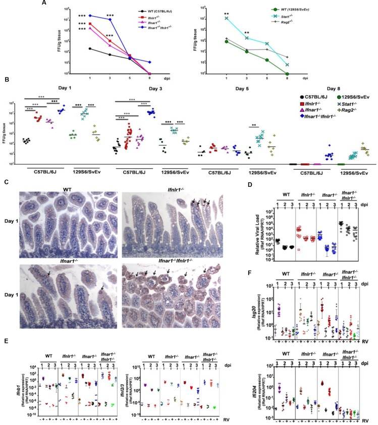
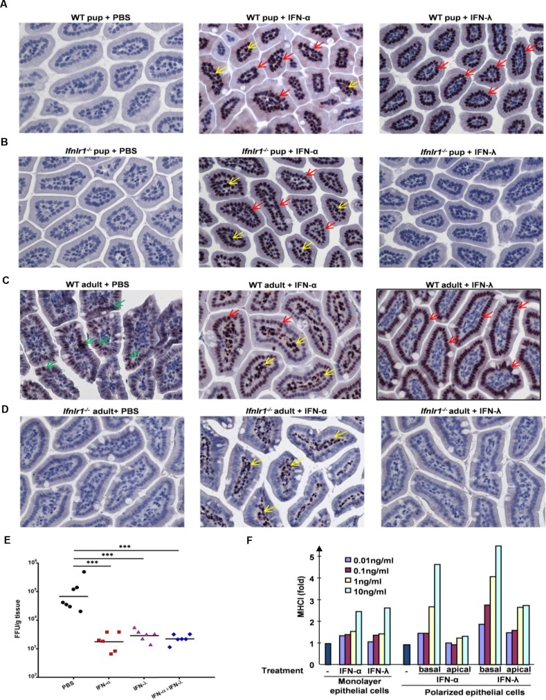
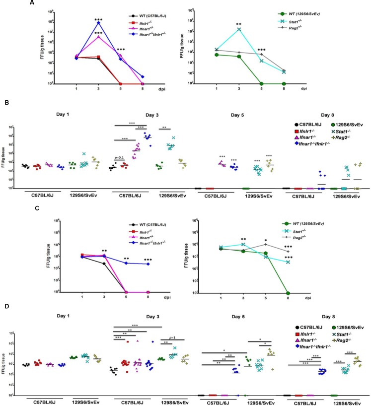
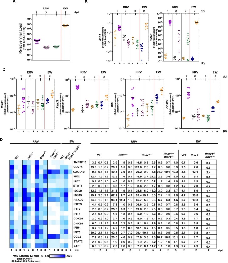
Type I (IFN-α/β) and type III (IFN-λ) interferons (IFNs) exert shared antiviral activities through distinct receptors. However, their relative importance for antiviral protection of different organ systems against specific viruses remains to be fully explored. We used mouse strains deficient in type-specific IFN signaling, STAT1 and Rag2 to dissect distinct and overlapping contributions of type I and type III IFNs to protection against homologous murine (EW-RV strain) and heterologous (non-murine) simian (RRV strain) rotavirus infections in suckling mice. Experiments demonstrated that murine EW-RV is insensitive to the action of both types of IFNs, and that timely viral clearance depends upon adaptive immune responses. In contrast, both type I and type III IFNs can control replication of the heterologous simian RRV in the gastrointestinal (GI) tract, and they cooperate to limit extra-intestinal simian RRV replication. Surprisingly, intestinal epithelial cells were sensitive to both IFN types in neonatal mice, although their responsiveness to type I, but not type III IFNs, diminished in adult mice, revealing an unexpected age-dependent change in specific contribution of type I versus type III IFNs to antiviral defenses in the GI tract. Transcriptional analysis revealed that intestinal antiviral responses to RV are triggered through either type of IFN receptor, and are greatly diminished when receptors for both IFN types are lacking. These results also demonstrate a murine host-specific resistance to IFN-mediated antiviral effects by murine EW-RV, but the retention of host efficacy through the cooperative action by type I and type III IFNs in restricting heterologous simian RRV growth and systemic replication in suckling mice. Collectively, our findings revealed a well-orchestrated spatial and temporal tuning of innate antiviral responses in the intestinal tract where two types of IFNs through distinct patterns of their expression and distinct but overlapping sets of target cells coordinately regulate antiviral defenses against heterologous or homologous rotaviruses with substantially different effectiveness.


Conflict of interest statement

I have read the journal's policy and the authors of this manuscript have the following competing interests: SVK is an inventor on patents and patent applications related to IFN-λs, which have been licensed for commercial development.

Figures

Fig 1
Fig 1. Generation and evaluation of IFN-λR1 KO mice.
(A) The exon-intron structure of the Ifnlr1 gene and the outline of the KO targeting vector are schematically shown. Exon 3 of the mouse Ifnlr1 gene was flanked by two loxp sites introduced into corresponding introns away from the splice signals. Remaining FRT site and positions of two primers (p1 and p2) are schematically mapped. (B) RNA samples were obtained from kidney cells of WT and Ifnlr1 -/- mice and used for RT-PCR with Ifnlr1-specific primers (p1 and p2) flanking exon 3. The sizes of the PCR fragments were determined by the agarose gel electrophoresis with 100 base pair DNA ladder shown in the left lane. (C) Sequencing of the PCR fragment from the Ifnlr1 -/- mice revealed splicing of exon 2 into exon 4 of the Ifnlr1 gene. A part of the Ifnlr1 gene transcript with original reading frame (black letters) and a shifted reading frame with a premature stop codon (orange letters) that was generated as a result of exon 3 skipping are shown. Arrows indicated positions of introns 1 through 4. (D) 8-day-old WT and Ifnlr1 -/- pups were intradermally injected with 1μg human IFN-αA/D (IFN-α) or murine IFN-λ2 (IFN-λ), 30 min later the indicated organs (small intestine (SI), large intestine (LI), lung, kidney and liver) were collected and analyzed for the presence of phosphorylated STAT1 (pSTAT1), as evidence of signaling through the IFN-αR or IFN-λR, by immunoblotting with pSTAT1-specific antibody. Immonoblotting with β actin antibody was used to evaluate equal loading.
Fig 2
Fig 2. Murine EW-RV efficiently replicates in intestine of suckling mice, regardless of the IFN action.
(A and B) Eight-day-old suckling WT (n = 6–13), Ifnar1 -/- (n = 7–11), Ifnlr1 -/- (n = 8–12), Ifnar1 -/- Ifnlr1 -/- (n = 8–12) mice (on C57BL/6J background) and WT (n = 11–16), Stat1 -/- (n = 13–16), Rag2 -/- (n = 6–8) mice (on 129S6/SvEv background) were orally infected with 104 DD50 EW-RV. Stool samples were collected daily from 2 to 12 dpi, and EW-RV fecal shedding determined by ELISA and expressed as OD unit. The variable sample number (n) reflects the variation of animals per time point. (A) Graph of kinetics of mean fecal EW-RV shedding in indicated strains of mice on C57BL/6J or 129S6/SvEv background. (B) Dot plot presentations of EW-RV shedding in indicated mouse strains on 3, 5 and 9 dpi. (C) Representative immunohistochemical staining of EW-RV in small intestine of WT and Stat1 -/- mice (on 129S6/SvEv background) on 1 dpi. (D-F) Graph of (D) quantitative RT-PCR detection of EW-RV levels, (E) IFN expression and (F) expression of indicated ISGs in small intestine of EW-RV-infected mice on 2 dpi. Each symbol (B and D-F) represents an individual mouse; horizontal lines indicate the mean (± SEM). *: significant difference (P < 0.05); **: significant difference (P < 0.01); ***: significant difference (P < 0.001).
Fig 3
Fig 3. IFNs may affect diarrheal disease but not weight gain during RV infection.
Eight-day-old suckling 129S6/SvEv, Stat1 -/- and Rag2 -/- (on 129S6/SvEv background) mice (n = 6–10) were orally infected with 104 DD50 EW-RV or 4x10⁶ FFU RRV. The variable sample number (n) reflects the variation of animals per time point. (A) Graph of average diarrheal disease and (B) average ratio of body weight between RV-infected and uninfected suckling mice were monitored daily in EW-RV (2 to 12 dpi) or RRV-infected suckling mice (2 to 8 dpi).
Fig 4
Fig 4. Both type I and type III IFNs contribute to intestinal antiviral immunity of suckling mice to simian RRV.
Eight-day-old suckling WT (n = 5–14), Ifnar1 -/- (n = 6–12), Ifnlr1 -/- (n = 8–20) and Ifnar1 -/- Ifnlr1 -/- mice (n = 6–10) (on C57BL/6J background), and WT (n = 6), Stat1 -/- (n = 6–8) and Rag2 -/- mice (n = 6) (on 129S6/SvEv background) were orally infected with 4x10⁶ FFU RRV. Small intestines were collected at indicated dpi and virus titers were determined by immunohistochemical infectious focus assay and expressed as FFU/g of tissue. The variable sample number (n) reflects the variation of animals per time point. (A) Graph of mean kinetics of RRV replication in small intestine of suckling mice of various strains on C57BL/6J or 129S6/SvEv background. (B) Dot plot presentations of intestinal RRV titers in suckling mice of indicated strains on 1, 3, 5 and 8 dpi. (C) Representative immunohistochemical staining of RRV antigens in small intestine of RRV-infected C57BL/6J WT, Ifnlr1 -/-, Ifnar1 -/- and Ifnar1 -/- Ifnlr1 -/- mice on 1 dpi. (D-F) Quantitative RT-PCR detection of (D) RRV levels, (E) IFN expression and (F) expression of indicated ISGs in small intestine of RRV-infected mice on 1, 2 and 3 dpi. Each symbol (B and D-F) represents an individual mouse; horizontal lines indicate the mean (± SEM). *: significant difference (P < 0.05); **: significant difference (P < 0.01); ***: significant difference (P < 0.001).
Fig 5
Fig 5. Differential responsiveness of IECs and LPCs to type I and type III IFNs.
(A-C) Representative of immunohistochemical staining of pSTAT1 (pTyr701) in small intestine of eight-day-old suckling (A) C57BL/6J WT or (B) Ifnlr1 -/- mice, or six to eight-week-old (C) C57BL/6J WT mice or (D) Ifnlr1 -/- mice were subcutaneously injected with PBS, human IFN-αA/D (IFN-α; 1μg) or murine IFN-λ2 (IFN-λ; 1μg). Small intestine samples were collected 30 min post injection. Red and yellow arrows indicate nuclear staining of pSTAT1 in IFN-treated IECs and LPCs, respectively. Green arrows indicate low levels of pSTAT1 staining in PBS-treated IECs in adult WT mice. (E) Graph of virus titers on 1 dpi in small intestine of eight-day-old suckling C57BL/6J WT mice subcutaneously injected with PBS, human IFN-αA/D (IFN-α; 1μg) or murine IFN-λ2 (IFN-λ; 1μg), or their combination (1μg of each IFN) 6 h before oral infection with 4x10⁶ FFU RRV. Virus titers were determined by immunohistochemical focus assay and expressed as FFU/g of tissue. (F) Graph of fold increase of MHC class I expression in human SW-1116 cells grown under polarized or regular culture conditions, treated at the apical or basolateral surfaces with various amounts of human IFN-αA/D or IFN-λ1 as indicated for 72 h. One representative experiment out of two is shown.
Fig 6
Fig 6. Type I IFNs control extra-intestinal spread and replication of simian RRV in MLN, but both IFN types limit simian RRV replication in liver.
Eight-day-old suckling WT (n = 6–8), Ifnar1 -/- (n = 6–13), Ifnlr1 -/- (n = 6–13) and Ifnar1 -/- Ifnlr1 -/- (n = 6–13) mice (on C57BL/6J background), and WT (n = 6), Stat1 -/- (n = 6–8) and Rag2 -/- mice (n = 6–8) (on 129S6/SvEv background) were orally infected with 4x10⁶ FFU RRV. MLN and liver samples were collected at indicated dpi and virus titers were determined by immunohistochemical infectious focus assay and expressed as FFU/g of tissue. The variable sample number (n) reflects the variation of animals per time point. (A and C) Graph of mean kinetics of RRV replication in MLN (A) and liver (C) of suckling mice of indicated strains on C57BL/6J or 129S6/SvEv background. (B and D) Dot plot presentations of RRV titers in MLN (B) and liver (D) of suckling mice of indicated strains on 1, 3, 5 and 8 dpi. Each symbol (B and D) represents an individual mouse; horizontal lines indicate the mean (± SEM).*: significant difference (P < 0.05); **: significant difference (P < 0.01); ***: significant difference (P < 0.001).
Fig 7
Fig 7. Type I and type III IFNs act redundantly during murine EW-RV and simian RRV infection to establish antiviral programs in small intestine.
(A-C) Graph of quantitative RT-PCR detection of (A) RV levels, (B) IFN expression and (C) expression of indicated ISGs in small intestine of RRV-infected WT mice on 1, 2 and 3 dpi and EW-RV-infected WT mice on 2 dpi. Symbols (A-C) duplicate measures from individual mice. (D) Graph of heat map of gene expression in intestinal samples presented as mean fold change in expression in infected mice relative to that of uninfected mice and shown on a 2-log scale. Transcriptional profiling was performed on small intestine samples from RRV and EW-RV-infected WT and single or double IFN receptor-deficient mice. Numbers in the table represent the mean fold change in gene expression on 1, 2 and 3 dpi for RRV, or on 2 dpi for EW-RV-infected, compared to uninfected litters. Changes above 3.0-fold are shown in bold. (n = 12–24 mice per group for RRV and 4–8 mice per group for EW-RV). Replication data in (A) is reformatted from Figs 2 and 4 for comparative purposes. Symbols (A-D) duplicate measures from individual mice. (n = 12–24 mice per group for RRV and 4–8 mice per group for EW-RV).

Similar articles

Cited by

References

    1. Ramig RF. Pathogenesis of intestinal and systemic rotavirus infection. J Virol. 2004;78(19):10213–20. Epub 2004/09/16. doi: 10.1128/JVI.78.19.10213–10220.2004 78/19/10213 [pii]. . - DOI - PMC - PubMed
    1. Tate JE, Burton AH, Boschi-Pinto C, Steele AD, Duque J, Parashar UD. 2008 estimate of worldwide rotavirus-associated mortality in children younger than 5 years before the introduction of universal rotavirus vaccination programmes: a systematic review and meta-analysis. Lancet Infect Dis. 2012;12(2):136–41. Epub 2011/10/28. doi: S1473-3099(11)70253-5 [pii] 10.1016/S1473-3099(11)70253-5 . - DOI - PubMed
    1. Mossel EC, Ramig RF. Rotavirus genome segment 7 (NSP3) is a determinant of extraintestinal spread in the neonatal mouse. J Virol. 2002;76(13):6502–9. Epub 2002/06/07. . - PMC - PubMed
    1. Mossel EC, Ramig RF. A lymphatic mechanism of rotavirus extraintestinal spread in the neonatal mouse. J Virol. 2003;77(22):12352–6. Epub 2003/10/29. . - PMC - PubMed
    1. Fenaux M, Cuadras MA, Feng N, Jaimes M, Greenberg HB. Extraintestinal spread and replication of a homologous EC rotavirus strain and a heterologous rhesus rotavirus in BALB/c mice. J Virol. 2006;80(11):5219–32. Epub 2006/05/16. doi: 80/11/5219 [pii] 10.1128/JVI.02664-05 . - DOI - PMC - PubMed

Publication types