. 2016 Jul 15;11(7):1827-33.
doi: 10.1021/acschembio.6b00324. Epub 2016 May 6.

Hepatitis C Virus Selectively Alters the Intracellular Localization of Desmosterol

Affiliations

Hepatitis C Virus Selectively Alters the Intracellular Localization of Desmosterol

Valerie A Villareal et al. ACS Chem Biol. .

Abstract

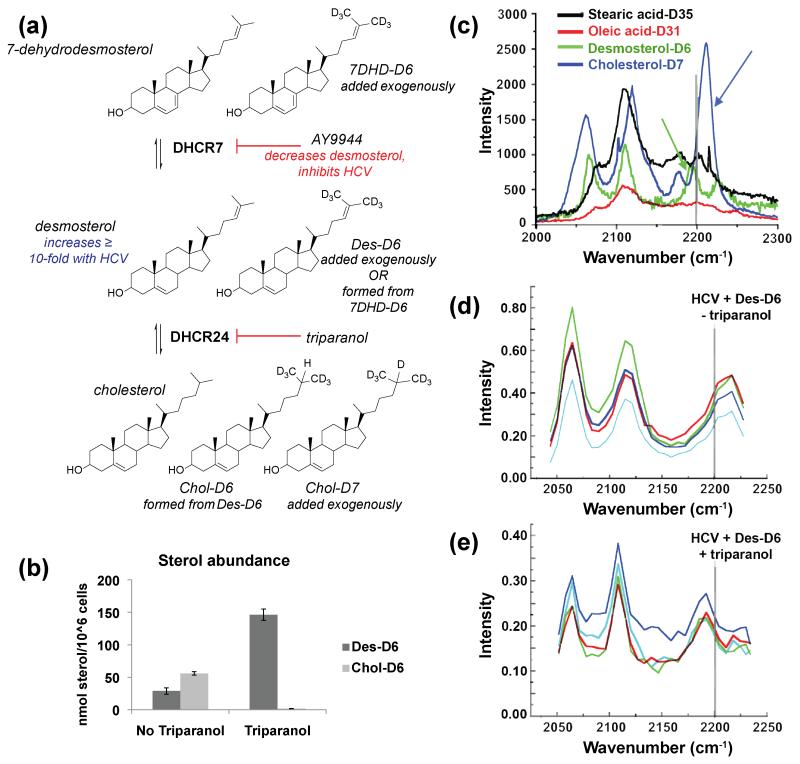
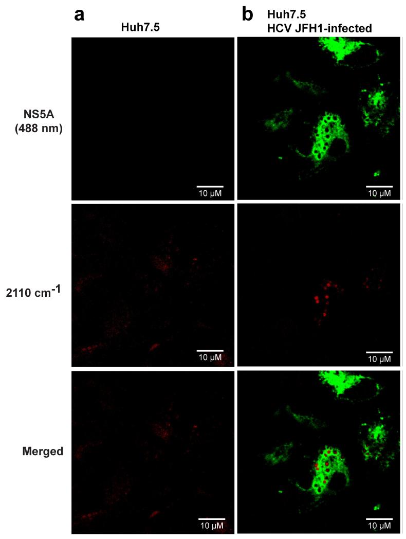
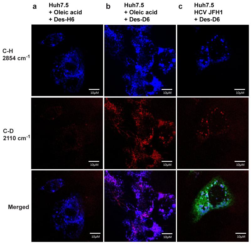
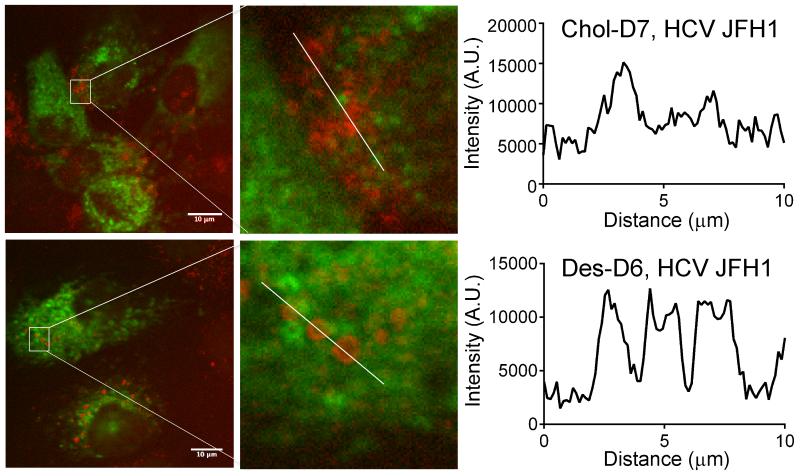
Hepatitis C virus (HCV) increases intracellular desmosterol without affecting the steady-state abundance of other sterols, and the antiviral activity of inhibitors of desmosterol synthesis is suppressed by the addition of exogenous desmosterol. These observations suggest a model in which desmosterol has a specific function, direct or indirect, in HCV replication and that HCV alters desmosterol homeostasis to promote viral replication. Here, we use stimulated Raman scattering (SRS) microscopy in combination with isotopically labeled sterols to show that HCV causes desmosterol to accumulate in lipid droplets that are closely associated with the viral NS5A protein and that are visually distinct from the broad distribution of desmosterol in mock-infected cells and the more heterogeneous and disperse lipid droplets to which cholesterol traffics. Localization of desmosterol in NS5A-associated lipid droplets suggests that desmosterol may affect HCV replication via a direct mechanism. We anticipate that SRS microscopy and similar approaches can provide much needed tools to study the localization of specific lipid molecules in cellulo and in vivo.


Figures

Figure 1
Figure 1. Conversion of exogenously added deuterated desmosterol to cholesterol is inhibited by triparanol
(a) Sterols and enzymes in late-stages of the Bloch pathway. HCV-JFH1 causes a ten-fold or greater increase in intracellular desmosterol. Deuterated substrates (Des-D6, 7DHD-D6, and Chol-D7) and inhibitors (AY9944, triparanol) are indicated. (b) LC-MS of extracted whole cell lipidomes. Lipids were extracted from Huh7.5 cells treated with triparanol and deuterated desmosterol (Des-D6). Whole-cell lipidomes were analyzed by LC-MS to quantify the abundance of Des-D6 and Chol-D6. Error bars represent the standard deviation of n = 2 biological replicates. (c-e) Hyperspectral SRS microscopy wave number scans of (c) deuterated standards collected under cell-free conditions and of individual puncta in HCV JFH1-infected Huh7.5 cells treated with (d) Des-D6 alone or (e) Des-D6 with triparanol. For (c), the deuterated cholesterol (Chol-D7) standard has a peak at wave number greater than 2200 nm-1 (blue arrow) whereas the deuterated desmosterol standard has a peak at a lower wave number, less than 2200 nm-1 (green arrow). For (d) and (e), each colored trace represents the wavelength scan of an individual punctum. The spectra obtained for puncta in HCV JFH1-infected cells treated with triparanol (e) is similar to the Raman spectrum of the desmosterol standard (c). For (c-e), each scan represents an analyzed punctum (n > 4).
Figure 2
Figure 2. Deuterated desmosterol forms distinct puncta in HCV-infected Huh7.5 cells
Huh7.5 cells were infected with HCV JFH1 or mock-infected and then treated with triparanol and exogenous deuterated desmosterol (Des-D6). 48 hours post-infection, cells were fixed, and immunofluorescence staining for the HCV NS5A protein was performed. SRS microscopy lasers were tuned to the C-D stretch (2110 cm−1) of deuterated desmosterol (Des-D6). Des-D6 and HCV viral protein, NS5A, are pseudocolored red and green, respectively. Representative images of (a) mock-infected and (b) HCV JFH1-infected Huh7.5 cells are presented from an experiment performed independently n ≥ 2. Additional representative images are provided in Fig. S3. Scale bar equals 10 micrometers.
Figure 3
Figure 3. Des-D6 colocalizes with lipid droplets in cells treated with oleic acid or infected with HCV JFH1
Huh7.5 cells were treated with oleic acid (a, b) or infected with HCV-JFH1 (c) to induce lipid droplet formation. Cells were treated with 2 μM triparanol and supplemented with exogenous Des-H6 (a) or Des-D6 (b, c). 48 hours post-infection, cells were fixed, and imaged. SRS microscopy lasers were tuned to the C-D stretch (2110 cm−1, pseudocolored red) of deuterated desmosterol (Des-D6) and to the C-H stretch (2854 cm−1, pseudocolored blue) present in endogenous lipids and also present in exogenously added Des-H6. Note that little or no signal is detected in cells supplemented with Des-H6 when the lasers are tuned to detect the C-D bond (2110 cm−1), demonstrating the selectivity of this imaging method for the C-D bond. Data shown are representative images from experiments that were independently performed n = 2 times. Scale bar equals 10 micrometers.
Figure 4
Figure 4. Des-D6 and Chol-D7 localized in lipid droplets with distinct visual phenotypes
Huh7.5 cells were treated with triparanol, infected with HCV-JFH1, and supplemented with Chol-D7 or Des-D6 as indicated. At 48 hours post-infection, cells were fixed and imaged by SRS microscopy to visualize localization of the C-D bond stretch at 2110 cm−1 for Des-D6 and 2220 cm−1 for Chol-D7. Chol-D7 localizes in puncta that are smaller and that co-localize in amorphous clusters when compared to Des-D6. The number of wavenumber stacks imaged for each sample were identical, and the representative images shown correspond to a single image made from identical stack numbers. Note, the stacks described represent shift in wavenumber (see Fig. 1c), and do not represent imaging in the z-plane. C-D intensity was plotted as function of distance in regions designated by white lines using ImageJ. Scale bar equals 10 micrometers.

References

    1. World Health Organization, M. C. Hepatitis C. Jul, 2015.
    1. Lonardo A, Adinolfi LE, Restivo L, Ballestri S, Romagnoli D, Baldelli E, Nascimbeni F, Loria P. Pathogenesis and significance of hepatitis C virus steatosis: an update on survival strategy of a successful pathogen. World J Gastroenterol. 2014;20:7089–7103. - PMC - PubMed
    1. Clejan S, Bittman R. Distribution and movement of sterols with different side chain structures between the two leaflets of the membrane bilayer of mycoplasma cells. J Biol Chem. 1984;259:449–455. - PubMed
    1. Nelson LD, Johnson AE, London E. How interaction of perfringolysin O with membranes is controlled by sterol structure, lipid structure, and physiological low pH: insights into the origin of perfringolysin O-lipid raft interaction. J Biol Chem. 2008;283:4632–4642. - PubMed
    1. Barba G, Harper F, Harada T, Kohara M, Goulinet S, Matsuura Y, Eder G, Schaff Z, Chapman MJ, Miyamura T, Brechot C. Hepatitis C virus core protein shows a cytoplasmic localization and associates to cellular lipid storage droplets. Proc Natl Acad Sci U S A. 1997;94:1200–1205. - PMC - PubMed

Publication types

LinkOut - more resources