. 2016 Apr 27;17(5):629.
doi: 10.3390/ijms17050629.

The Glutathione Derivative, GSH Monoethyl Ester, May Effectively Whiten Skin but GSH Does Not

Affiliations

The Glutathione Derivative, GSH Monoethyl Ester, May Effectively Whiten Skin but GSH Does Not

Bo Young Chung et al. Int J Mol Sci. .

Abstract

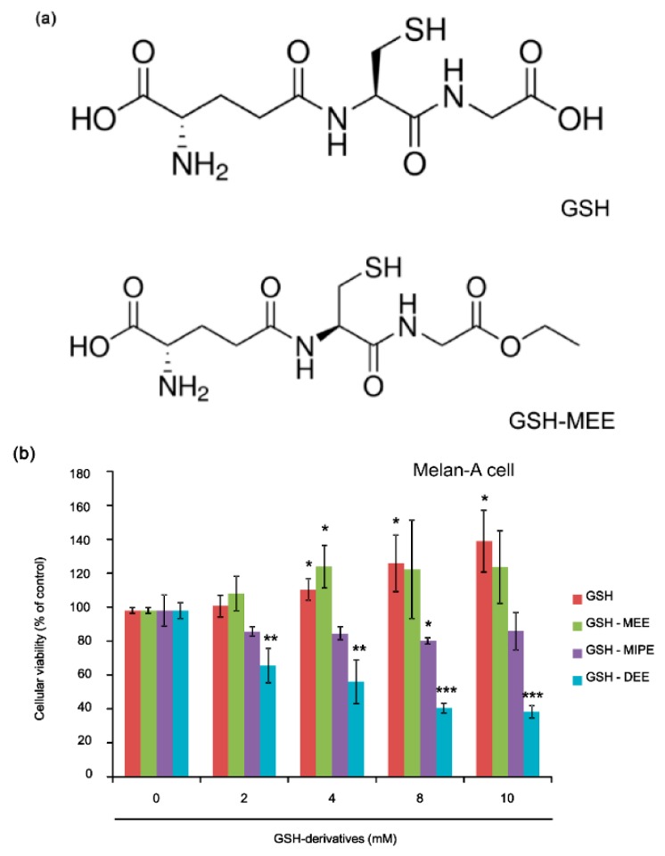
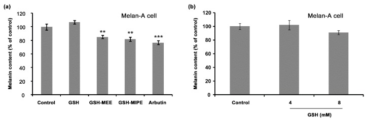
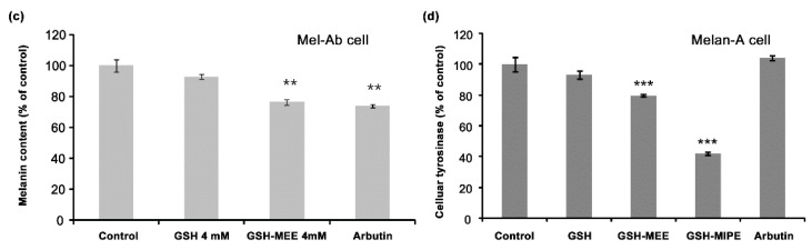
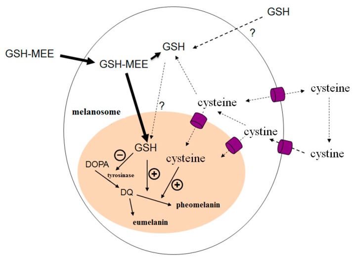
Glutathione in its reduced form (GSH) is an antioxidant and also is involved in pheomelanin formation. Thus, it has been long believed that GSH has a skin whitening effect. However, its actual or direct effect is unproven. We evaluated the anti-melanogenic effects of GSH and its derivatives in vitro. We examined change of melanogenesis and its related proteins by GSH itself and its derivatives, including GSH monoethyl ester (GSH-MEE), GSH diethyl ester (GSH-DEE) and GSH monoisopropyl ester (GSH-MIPE) in Melan-A cells, Mel-Ab cells, and B16F10 cells. GSH and GSH-MEE did not display cytotoxic activity, but GSH-MIPE and GSH-DEE did. Intriguingly, GSH itself had no inhibitory effect on melanin production or intracellular tyrosinase activity. Rather, it was GSH-MEE and GSH-MIPE that profoundly reduced the amount of melanin and intracellular tyrosinase activity. Thus, GSH-MEE was selected as a suitable candidate skin-whitening agent and it did not alter melanogenesis-associated proteins such as microphthalmia-associated transcription factor (MITF), tyrosinase, tyrosinase-related protein (TRP)-1, and TRP-2, but it did increase the amount of suggested pheomelanin and suggested pheomelanin/eumelanin ratio. GSH-MEE was effective for anti-melanogenesis, whereas GSH itself was not. GSH-MEE could be developed as a safe and efficient agent for the treatment of hyperpigmentation skin disorders.

Keywords: glutathione derivatives; glutathione monoethyl ester; melanogenesis; pheomelanin.


Figures

Figure 1
Figure 1
(a) Chemical structures of reduced glutathione (GSH) and GSH monoethyl ester (GSH-MEE); (b) Effect of GSH derivatives on cellular viability. GSH and GSH-MEE had no effect on cellular viability, but GSH-MIPE and GSH-DEE showed toxicity in Melan-A cells. Data are shown as the mean ± SD of three replicates, and * p < 0.05, ** p < 0.01, and *** p < 0.001 compared to untreated controls. GSH, reduced glutathione; GSH-MEE, GSH monoethyl ester; GSH-MIPE, GSH monoisopropyl ester; GSH-DEE, GSH diethyl ester; SD, standard deviation.
Figure 2
Figure 2
(a) Effect of GSH derivatives on melanin content in Melan-A cells. GSH exhibited no inhibitory effect on melanin production, but GSH-MEE and GSH-MIPE decreased the melanin synthesis in Melan-A cells. Arbutin (100 µg/mL) was treated to serve as a positive control. The melanin content was calculated by normalizing the melanin content to total cellular protein and reported as a percentage of control. Data are shown as the mean ± SD of three replicates, and ** p < 0.01, and *** p < 0.001. GSH, reduced glutathione; GSH-MEE, GSH monoethyl ester; GSH-MIPE, GSH monoisopropyl ester; SD, standard deviation; (b) The effect of GSH on melanin production in Melan-A cells. GSH had no inhibitory effect on melanin production in Melan-A cells. Each sample was quantified with the same amount of protein and reported as a percentage of control. Data are shown as the mean ± SD of three replicates; (c) Effect of GSH and GSH-MEE on melanin content in Mel-Ab cells GSH had no inhibitory effect on melanin production, but GSH-MEE decreased the synthesis of melanin significantly in Mel-Ab cells. Arbutin (100 µg/mL) was treated as a positive control. The melanin content was calculated by normalizing the melanin content to total cellular protein and reported it as a percentage of control. Data are shown as the mean ± SD of three replicates, and ** p < 0.01; (d) the effect of GSH derivatives on intracellular tyrosinase activity in Melan-A cells. GSH had no significant effect on intracellular tyrosinase activity, but GSH-MEE and GSH-MIPE decreased the activity of intracellular tyrosinase in Melan-A cells. Arbutin (100 µg/mL) was treated as a positive control. Each sample was quantified with the same amount of protein and reported as a percentage of control. Data are shown as the mean ± SD of three replicates, and *** p < 0.001.
Figure 2
Figure 2
(a) Effect of GSH derivatives on melanin content in Melan-A cells. GSH exhibited no inhibitory effect on melanin production, but GSH-MEE and GSH-MIPE decreased the melanin synthesis in Melan-A cells. Arbutin (100 µg/mL) was treated to serve as a positive control. The melanin content was calculated by normalizing the melanin content to total cellular protein and reported as a percentage of control. Data are shown as the mean ± SD of three replicates, and ** p < 0.01, and *** p < 0.001. GSH, reduced glutathione; GSH-MEE, GSH monoethyl ester; GSH-MIPE, GSH monoisopropyl ester; SD, standard deviation; (b) The effect of GSH on melanin production in Melan-A cells. GSH had no inhibitory effect on melanin production in Melan-A cells. Each sample was quantified with the same amount of protein and reported as a percentage of control. Data are shown as the mean ± SD of three replicates; (c) Effect of GSH and GSH-MEE on melanin content in Mel-Ab cells GSH had no inhibitory effect on melanin production, but GSH-MEE decreased the synthesis of melanin significantly in Mel-Ab cells. Arbutin (100 µg/mL) was treated as a positive control. The melanin content was calculated by normalizing the melanin content to total cellular protein and reported it as a percentage of control. Data are shown as the mean ± SD of three replicates, and ** p < 0.01; (d) the effect of GSH derivatives on intracellular tyrosinase activity in Melan-A cells. GSH had no significant effect on intracellular tyrosinase activity, but GSH-MEE and GSH-MIPE decreased the activity of intracellular tyrosinase in Melan-A cells. Arbutin (100 µg/mL) was treated as a positive control. Each sample was quantified with the same amount of protein and reported as a percentage of control. Data are shown as the mean ± SD of three replicates, and *** p < 0.001.
Figure 3
Figure 3
(a) Effect of GSH-MEE on melanin production in Melan-A cells. GSH-MEE decreased the production of melanin in a dose-dependent manner in Melan-A cells. Arbutin (100 µg/mL) was treated as a positive control. The melanin content was calculated by normalizing the melanin content to total cellular protein and reported as a percentage of control. Data are shown as the mean ± SD of three replicates, and * p < 0.05, ** p < 0.01. GSH-MEE, reduced glutathione monoethyl ester; SD, standard deviation; (b) Effect of GSH-MEE on melanin content in B16F10 cells. GSH-MEE decreased the amount of melanin produced by B16F10 cells. Arbutin (100 µg/mL) was treated as a positive control. The melanin content was calculated by normalizing the melanin contents to total cellular protein and reported as a percentage of control. Data are shown as the mean ± SD of three replicates, and *** p < 0.001. GSH-MEE, reduced glutathione monoethyl ester; α-MSH, α-melanocyte-stimulating hormone; SD, standard deviation; (c) Effect of GSH-MEE on intracellular tyrosinase activity in Melan-A cells. GSH-MEE decreased the activity of intracellular tyrosinase in a dose-dependent manner in Melan-A cells. Arbutin (100 µg/mL) was treated as a positive control. Each sample was quantified with the same amount of protein and reported as a percentage of control. Data are shown as the mean ± SD of three replicates, and ** p < 0.01, and *** p < 0.001. GSH-MEE, reduced glutathione monoethyl ester; SD, standard deviation; (d) Effect of GSH-MEE on intracellular tyrosinase activity in B16F10 cells. GSH-MEE decreased the activity of intracellular tyrosinase in B16F10 cells. Arbutin (100 µg/mL) was treated as a positive control. Each sample was quantified with the same amount of protein and reported as a percentage of control. Data are shown as the mean ± SD of three replicates, and *** p < 0.001. GSH-MEE, reduced glutathione monoethyl ester; α-MSH, α-melanocyte-stimulating hormone; SD, standard deviation; (e) Effect of GSH-MEE on the expression of melanogenesis-related proteins. When Melan-A cells were treated with GSH-MEE, the expression levels of MITF, tyrosinase, TRP-1, and TRP-2 did not change significantly. Arbutin (100 µg/mL) was treated as a positive control, and β‑actin expression was used as the loading control. These results represents three independent experiments. GSH-MEE, reduced glutathione monoethyl ester; MITF, microphthalmia-associated transcription factor; TRP-1, tyrosinase-related protein-1; TRP-2, tyrosinase-related protein-2; (f) Effect of GSH-MEE on suggested eumelanin and pheomelanin production. When Melan-A cells were treated with GSH-MEE, the synthesis of melanin with absorbance at 400 nm (A400, suggestive of eumelanin) increased, whereas the production of melanin with absorbance at 350 (A350, suggestive of pheomelanin) decreased. Arbutin (100 µg/mL) was treated as a positive control. The melanin content was calculated by normalizing the melanin contents to total cellular proteins and reported as a percentage of control. Data are shown as the mean ± SD of three replicates, and * p < 0.05, ** p < 0.01, and *** p < 0.001. GSH-MEE, reduced glutathione monoethyl ester; A400, absorbance at 400 nm; A350, absorbance at 350 nm; SD, standard deviation.
Figure 4
Figure 4
Possible effects of GSH-MEE, GSH, and cysteine on melanogenesis. GSH synthesize intracellularly and exported to extracellular space. Cellular uptake of GSH itself is not probable. GSH-MEE cross the biological membranes by simple diffusion and metabolize intracellularly to generate GSH. Cysteine is readily transported across cell membranes but potentially toxic at high concentrations due to generation of reactive oxygen species and depletion of pyridoxal phosphate. GSH-MEE (but not GSH itself) could suppress the melanin production and tyrosinase activity and raise the suggested pheomelanin/eumelanin ratio. black arrows, melanin synthesis process flow; dotted black arrows, transport process flows of GSH, cysteine, and cysteine, bold black arrows, transport process flow of GSH-MEE; question marks, not yet confirmed; +, positive effects (activation); −, negative effects (suppression); purple shape, membrane channel. GSH, reduced glutathione; GSH-MEE, GSH monoethyl ester; DOPA, dihydroxyphenylalanine; DQ, dopaquinone.

Similar articles

Cited by

References

    1. Arjinpathana N., Asawanonda P. Glutathione as an oral whitening agent: A randomized, double-blind, placebo-controlled study. J. Dermatol. Treat. 2012;23:97–102. doi: 10.3109/09546631003801619. - DOI - PubMed
    1. Slominski A.T., Zmijewski M.A., Skobowiat C., Zbytek B., Slominski R.M., Steketee J.D. Sensing the environment: Regulation of local and global homeostasis by the skin’s neuroendocrine system. Adv. Anat. Embryol. Cell Biol. 2012;212:1–115. - PMC - PubMed
    1. Slominski A., Tobin D.J., Shibahara S., Wortsman J. Melanin pigmentation in mammalian skin and its hormonal regulation. Physiol. Rev. 2004;84:1155–1228. doi: 10.1152/physrev.00044.2003. - DOI - PubMed
    1. Slominski A., Zmijewski M.A., Pawelek J. l-tyrosine and l-dihydroxyphenylalanine as hormone-like regulators of melanocyte functions. Pigment Cell Melanoma Res. 2012;25:14–27. doi: 10.1111/j.1755-148X.2011.00898.x. - DOI - PMC - PubMed
    1. Slominski R.M., Zmijewski M.A., Slominski A.T. The role of melanin pigment in melanoma. Exp. Dermatol. 2015;24:258–259. doi: 10.1111/exd.12618. - DOI - PMC - PubMed

LinkOut - more resources