. 2016 May 31;7(22):32707-22.
doi: 10.18632/oncotarget.9017.

Extracellular miR-1246 promotes lung cancer cell proliferation and enhances radioresistance by directly targeting DR5

Affiliations

Extracellular miR-1246 promotes lung cancer cell proliferation and enhances radioresistance by directly targeting DR5

Dexiao Yuan et al. Oncotarget. .

Abstract

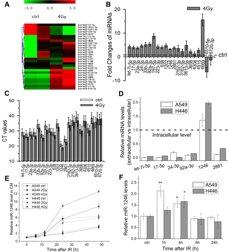
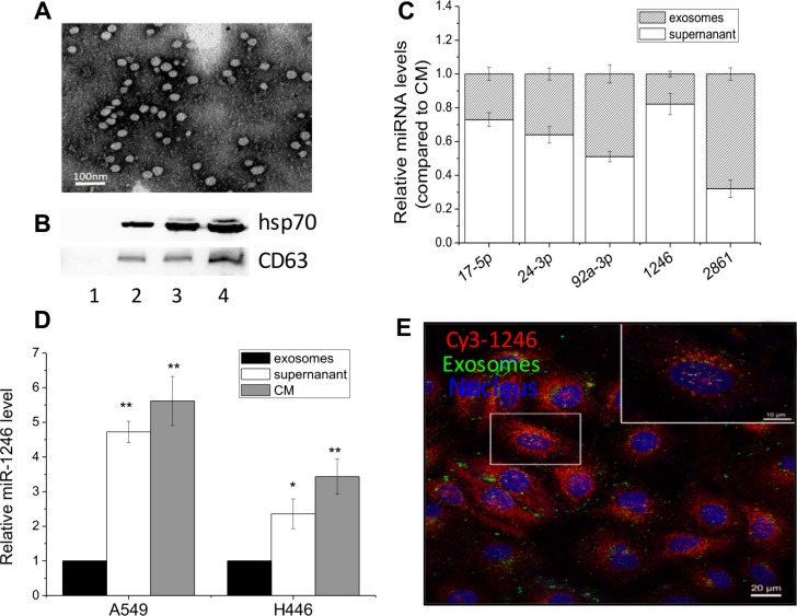
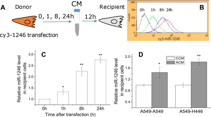
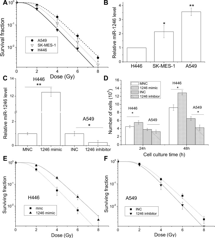
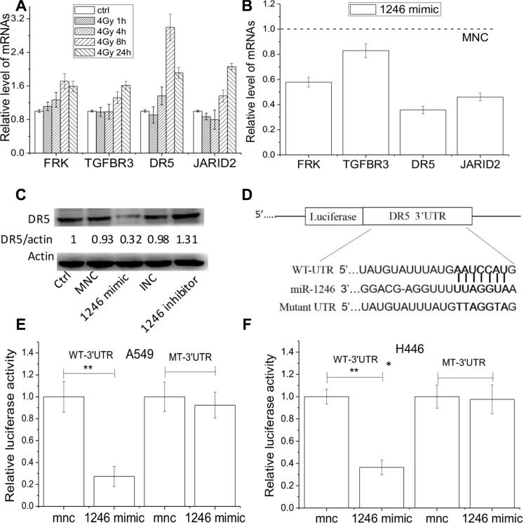
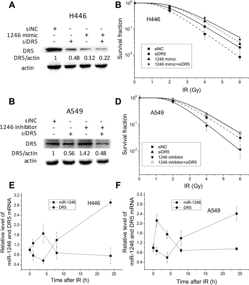
MiRNAs in the circulation have been demonstrated to be a type of signaling molecule involved in intercellular communication but little is known about their role in regulating radiosensitivity. This study aims to investigate the effects of extracellular miRNAs induced by ionizing radiation (IR) on cell proliferation and radiosensitivity. The miRNAs in the conditioned medium (CM) from irradiated and non-irradiated A549 lung cancer cells were compared using a microarray assay and the profiles of 21 miRNAs up and down-regulated by radiation were confirmed by qRT-PCR. One of these miRNAs, miR-1246, was especially abundant outside the cells and had a much higher level compared with that inside of cells. The expressions of miR-1246 in both A549 and H446 cells increased along with irradiation dose and the time post-irradiation. By labeling exosomes and miR-1246 with different fluorescence dyes, it was found that the extracellular miR-1246 could shuttle from its donor cells to other recipient cells by a non-exosome associated pathway. Moreover, the treatments of cells with miR-1246 mimic or its antisense inhibitor showed that the extracellular miR-1246 could enhance the proliferation and radioresistance of lung cancer cells. A luciferase reporter-gene transfer experiment demonstrated that the death receptor 5 (DR5) was the direct target of miR-1246, and the kinetics of DR5 expression was opposite to that of miR-1246 in the irradiated cells. Our results show that the oncogene-like extracellular miR-1246 could act as a signaling messenger between irradiated and non-irradiated cells, more importantly, it contributes to cell radioresistance by directly suppressing the DR5 gene.

Keywords: DR5 gene; bystander effect; exosomes; extracellular mR-1246; radioresistance.


Conflict of interest statement

The authors declare that there are no conflicts of interest in work described manuscript.

Figures

Figure 1
Figure 1. Radiation-induced alterations of extracellular miRNA expression profiles in lung cancer cells
(A) Hierarchical clustering of differentially expressed miRNAs in the CM of A549 cells at 24 h after 0 Gy or 4 Gy X-ray irradiation. Red indicates higher expression, green indicates lower expression, and black means no expression difference. (B) Quantification of 21 differentially miRNAs in the above CM confirmed by qRT-PCR. Results are expressed as relative levels compared with non-irradiated controls and normalized to spike control (cel-miR-39). Each reaction was performed in triplicate. (C) The CT values of above 21 differentially miRNAs with qRT-PCR assay. (D) Ratio of extracellular miRNAs to intracellular miRNAs of A549 and H446 cells. Equal amounts of RNAs (20 ng) isolated from cells and CM were analyzed with qRT-PCR. (E) Relative levels of extracellular miR-1246 in the CM of A549 and H446 cells after indicated times (0, 6, 12, 24 or 48 h) of irradiation (0, 2, 4 Gy). (F) Relative expression levels of intracellular miR-1246 in A549 and H446 cells 1, 4, 8 or 24 h after 4 Gy irradiation. Data were presented as means ± SEMs of three independent experiments. *P < 0.05, **P < 0.01 compared to non-irradiated controls.
Figure 2
Figure 2. Extracellular miR-1246 exists in non-exosomes associated form
(A) Exosomes released from A549 cells were observed under an electron microscopy. (B) Hsp70 and CD63 protein expressions in the exosomes purified from the medium with depleted exosomes FBS (lane 1) and the CM of A549 cells with a number of 2.5 × 10⁶ (lane 2), 5 × 10⁶ (lane 3), and 1 × 10⁷ (lane 4). (C) Comparison the levels of five miRNAs in the exosome-free supernatant and exosome-enriched pellet of the CM from A549 cells. The total expression level of each miRNA was set to 1. (D) Relative miR-1246 levels in the exosomes, exosome-free supernatant and the CM from A549 and H446 cells. The miR-1246 level in exosomes was set as control. Data were presented as means ± SEMs of five independent experiments (*P < 0.05,** P < 0.01). (E) Localization of exosomes and extracellular miR-1246 in the recipient A549 cells. The CM was collected from A549 cells labeled with DiO and Cy3-labeled miR-1246 and was then used to incubate other recipient A549 cells for 24 h. The recipient A549 cells with red fluorescence Cy3-labeled miR-1246, green fluorescence exosomes, and DAPI stained nuclear with blue fluorescence were fixed and observed under a confocal microscopy.
Figure 3
Figure 3. Extracellular miR-1246 was integrated into recipient cells
(A) The scheme of experiments. A549 cells were transfected with Cy3-labeled miR-1246, then the CM was collected at 0, 1, 8 and 24 h after transfection and used to culture other recipient A549 cells for 12 h. The uptake of miR-1246 by A549 cells was detected by flow cytometry (B) and qRT–PCR (C), respectively. (D) Relative levels of intracellular miR-1246 in A549 and H446 cells treated with CM from non-irradiated A549 cells (CCM) or from 4 Gy X-ray irradiated A549 cells (ACM) for 12 h. The expression level of miR-1246 in the cells treated with CCM was set as control. Data were presented as means ± SEMs of five independent experiments (*P < 0.05, **P < 0.01).
Figure 4
Figure 4. miR-1246 mimic promotes cell proliferation and enhances radioresistance
Lung cancer cell lines of A549, SK-MES-1 and H446 were irradiated with X-rays. (A) Dose response of cell survival. (B) Intracellular miR-1246 expressions of lung cancer cells. (C) The miR-1246 expressions in H446 and A549 cells transfected with miR-1246 mimic and its negative control (MNC), miR-1246 antisense inhibitor and its negative control (INC), respectively. (D) Cell number of above H446 and A549 cells at 24 and 48 h after transfection. (E) Clonogenic survivals of H446 cells transfected with miR-1246 mimic and its control. (F) Clonogenic survivals of A549 cells transfected with miR-1246 inhibitor and its control. Data were presented as means ± SEMs of four independent experiments (*P < 0.05, **P < 0.01).
Figure 5
Figure 5. DR5 is the direct target of miR-1246
Expressions of mRNA and protein were detected by RT-pCR and Western blot assay, respectively. (A) Relative expression levels of JARID2, FRK, DR5 and TGFBR3 in H446 cells after 1, 4, 8, and 24 h of 4 Gy irradiation. (B) Relative levels of the above four genes in H446 cells transfected with miR246 mimic in comparison of MNC. (C) DR5 protein expressions in H446 cells treated with miR-1246 mimic, inhibitor, or negative controls. Actin was used as an internal reference. (D) The predicted miR-1246 binding sites in the wild-type (WT) DR5 UTR and mutant DR5 UTR. (E, F) WT or mutant 3′UTR- DR5 Gaussian Luciferase reporter vector was co-transfected with miR-1246 mimic or its control into A549 cells and H446 cells, and Gaussian luciferase activity of each sample was normalized to the secreted alkaline phosphatase (SEAP). Data were presented as means ± SEMs of four independent experiments (*P < 0.05, **P < 0.01).
Figure 6
Figure 6. miR-1246 increases cancer cell radioresistance via directly targeting DR5
(A) DR5 protein expressions in the H446 cells transfected with miR-1246 mimic, DR5 siRNA and their negative controls, which were normalized to siRNA control with actin as an internal reference. (B) Dose response of the survival of H446 cells transfected with miR-1246 mimic, DR5 siRNA and their negative controls. (C) DR5 protein expressions in the A549 cells transfected with miR-1246 inhibitor, DR5 siRNA and their negative controls, which were normalized to siRNA control with actin as an internal reference. (D) Dose response of the survival of A549 cells transfected with miR-1246 inhibitor, DR5 siRNA and their negative controls. (E, F) The kinetics of miR-1246 and DR5 expressions in H446 and A549 cells after 4 Gy irradiation. Data were presented as means ± SEMs of four independent experiments.
Figure 7
Figure 7. Extracellular miR-1246 promotes cell proliferation and induces radioresistance via directly suppressing DR5 in bystander cells
(A) The scheme of experiments. A549 cells were irradiated with 4 Gy X-rays or transfected with miR-1246 mimics, miR-1246 inhibitor and their control, respectively. Then its CM was collected to treat non-irradiated bystander cells for 48 h. (B) miR-1246 expressions in the bystander H446 cells treated with the CM from A549 cells as shown in Figure 7A. (C) Proliferations of H446 and A549 cells after 48 h of incubation with the active CM (ACM) from A549 cells or its control (CCM). (D) Clonogenic survival of irradiated H446 cells that were treated with the CM indicated above. (E) DR5 protein expression in the bystander H446 cells that were treated with the CM indicated above. Data were presented as means ± SEMs of three independent experiments (*P < 0.05, **P < 0.01).

Similar articles

Cited by

References

    1. Willers H, Azzoli CG, Santivasi WL, Xia F. Basic mechanisms of therapeutic resistance to radiation and chemotherapy in lung cancer. Cancer J. 2013;19:200–207. - PMC - PubMed
    1. Barker HE, Paget JT, Khan AA, Harrington KJ. The tumour microenvironment after radiotherapy: mechanisms of resistance and recurrence. Nat Rev Cancer. 2015;15:409–425. - PMC - PubMed
    1. Zhao LQ, Bode AM, Cao Y, Dong ZG. Regulatory mechanisms and clinical perspectives of miRNA in tumor radiosensitivity. Carcinogenesis. 2012;33:2220–2227. - PMC - PubMed
    1. Mitchell PS, Parkin RK, Kroh EM, Fritz BR, Wyman SK, Pogosova-Agadjanyan EL, Peterson A, Noteboom J, O'Briant KC, Allen A, Lin DW, Urban N, Drescher CW, et al. Circulating microRNAs as stable blood-based markers for cancer detection. P Natl Acad Sci USA. 2008;105:10513–10518. - PMC - PubMed
    1. Weber JA, Baxter DH, Zhang SL, Huang DY, Huang KH, Lee MJ, Galas DJ, Wang K. The MicroRNA Spectrum in 12 Body Fluids. Clin Chem. 2010;56:1733–1741. - PMC - PubMed

MeSH terms

Substances