. 2016 Jun 10;291(24):12658-12672.
doi: 10.1074/jbc.M115.711325. Epub 2016 Apr 25.

Structural Analysis of a Complex between Small Ubiquitin-like Modifier 1 (SUMO1) and the ZZ Domain of CREB-binding Protein (CBP/p300) Reveals a New Interaction Surface on SUMO

Affiliations

Structural Analysis of a Complex between Small Ubiquitin-like Modifier 1 (SUMO1) and the ZZ Domain of CREB-binding Protein (CBP/p300) Reveals a New Interaction Surface on SUMO

Carl Diehl et al. J Biol Chem. .

Abstract

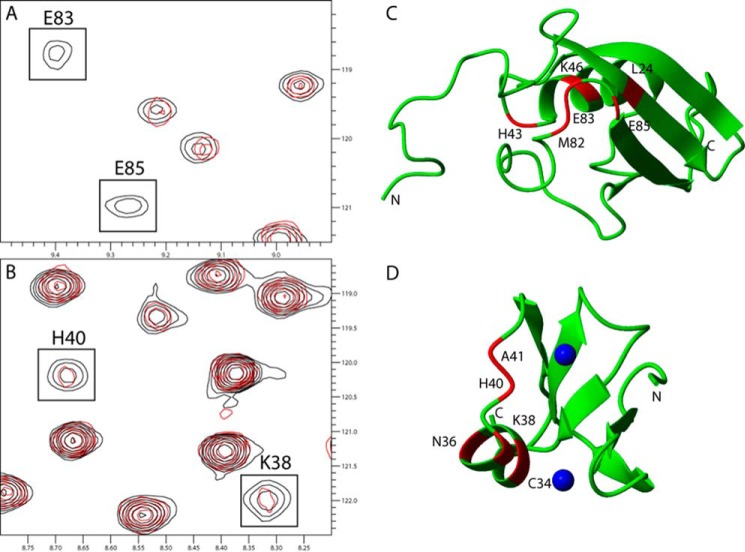
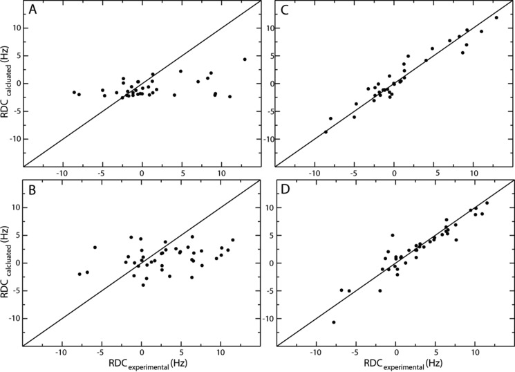
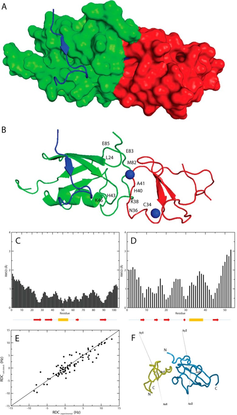
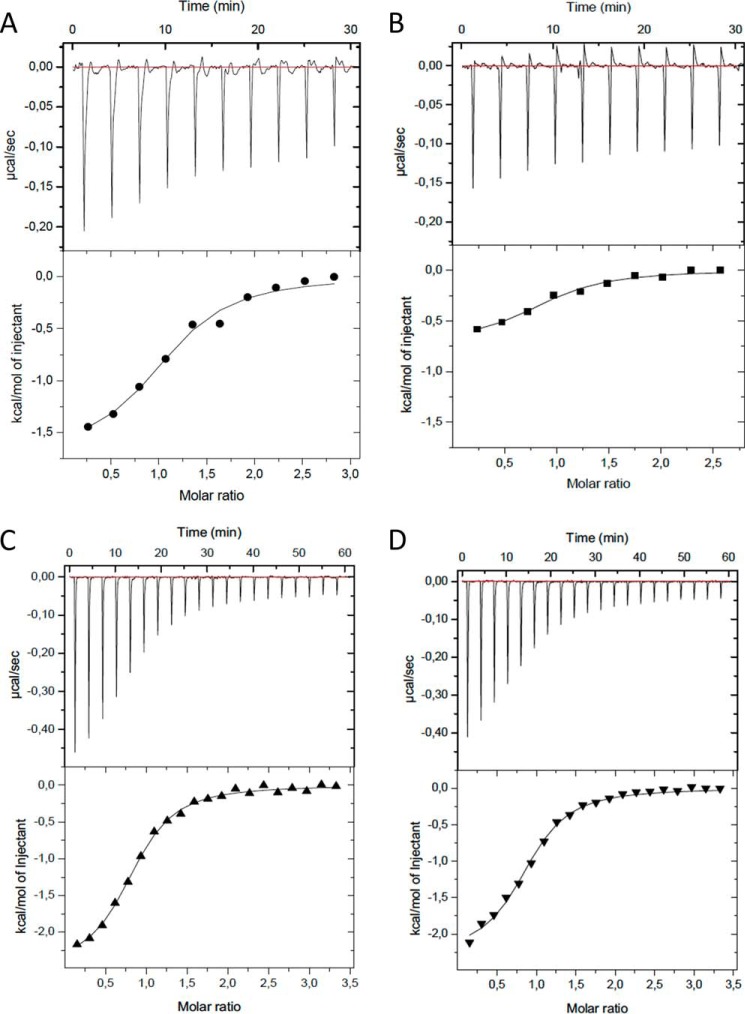
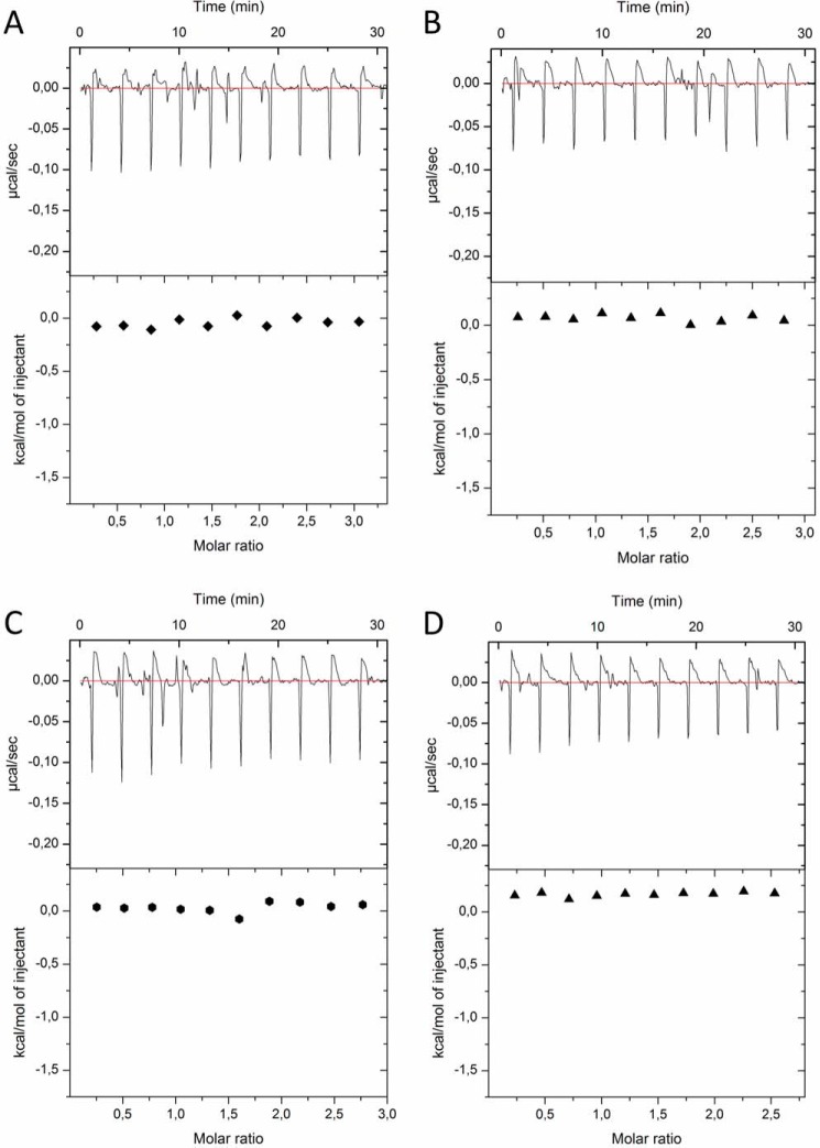
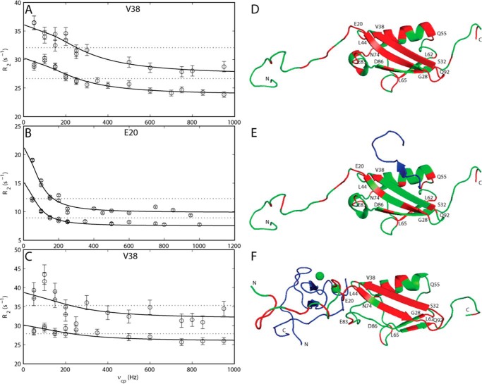
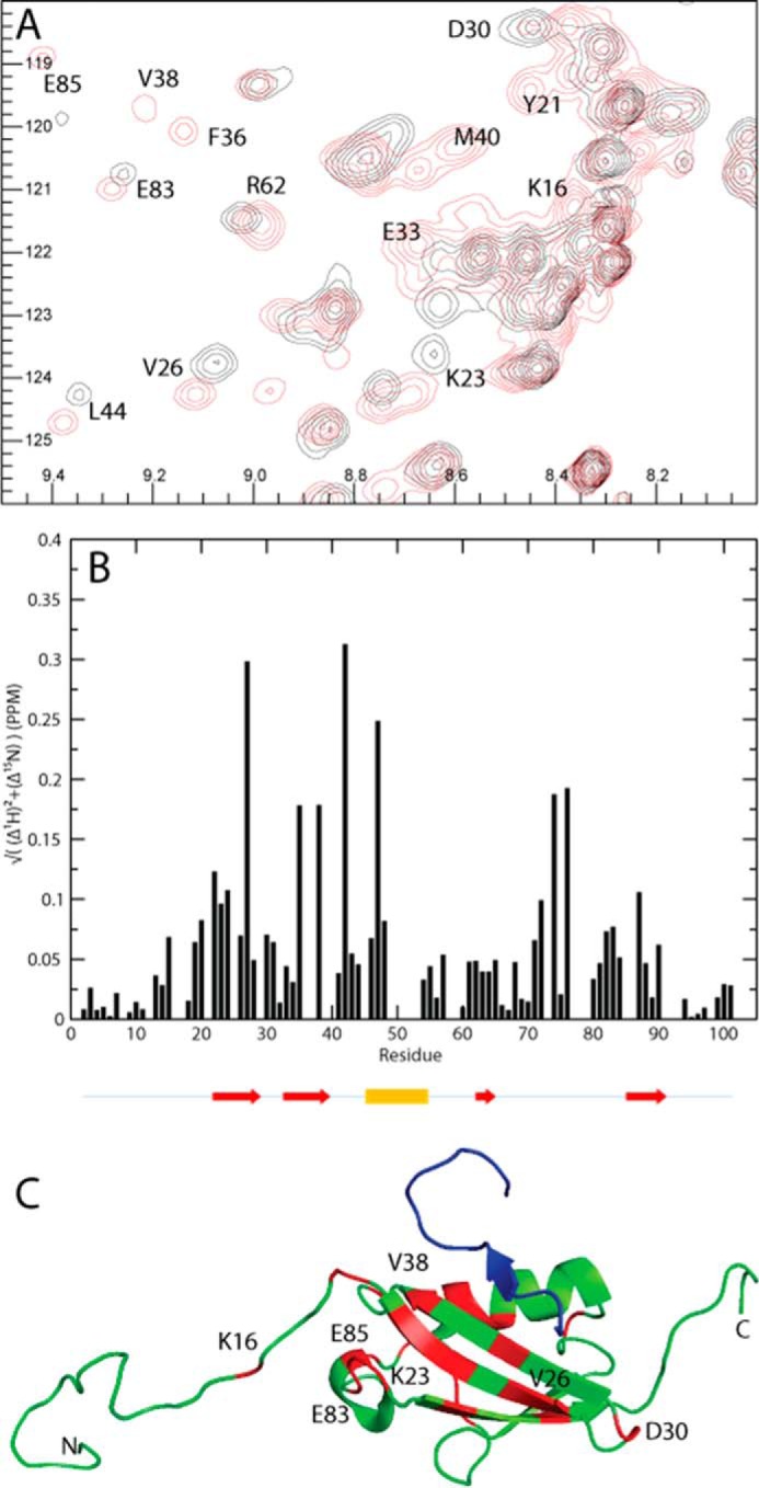
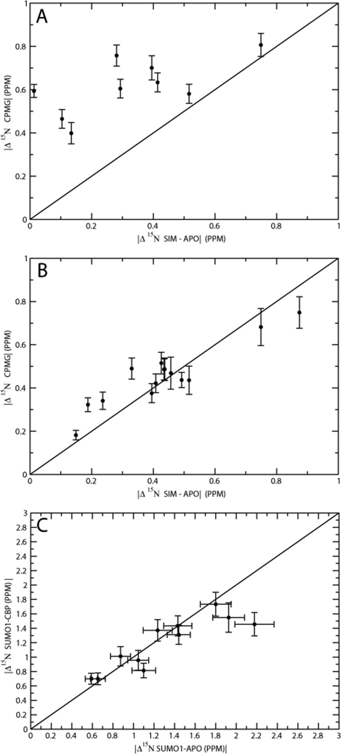
We have recently discovered that the ZZ zinc finger domain represents a novel small ubiquitin-like modifier (SUMO) binding motif. In this study we identify the binding epitopes in the ZZ domain of CBP (CREB-binding protein) and SUMO1 using NMR spectroscopy. The binding site on SUMO1 represents a unique epitope for SUMO interaction spatially opposite to that observed for canonical SUMO interaction motifs (SIMs). HADDOCK docking simulations using chemical shift perturbations and residual dipolar couplings was employed to obtain a structural model for the ZZ domain-SUMO1 complex. Isothermal titration calorimetry experiments support this model by showing that the mutation of key residues in the binding site abolishes binding and that SUMO1 can simultaneously and non-cooperatively bind both the ZZ domain and a canonical SIM motif. The binding dynamics of SUMO1 was further characterized using (15)N Carr-Purcell-Meiboom-Gill (CPMG) relaxation dispersions, which define the off rates for the ZZ domain and SIM motif and show that the dynamic binding process has different characteristics for the two cases. Furthermore, in the absence of bound ligands SUMO1 transiently samples a high energy conformation, which might be involved in ligand binding.

Keywords: SUMO-interacting motif (SIM); biophysics; nuclear magnetic resonance (NMR); protein dynamics; protein structure; protein-protein interaction; structural biology; structural model.


Figures

FIGURE 1.
FIGURE 1.
Chemical shift differences observed when titrating the unlabeled ZZ domain or SUMO1 into 13C,15N-labeled SUMO1 or ZZ domain, respectively. A, cutout of 15N HSQC showing backbone amides in SUMO1 affected by the binding of the ZZ domain. B, cutout of 15N HSQC showing backbone amides in the ZZ domain affected by the binding of SUMO1. Contours colored in black correspond to the apo form, whereas contours colored in red correspond to ZZ domain-bound SUMO1. C, residues in SUMO1 affected by the binding of ZZ are highlighted in red: Leu-24, His-43, Lys-46, Met-82, Glu-83, and Glu-85 (Protein Data Bank code 1A5R). D, residues in the ZZ domain affected by the binding of SUMO1 are highlighted in red: Cys-34, Asn-36, Lys-38, His-40, and Ala-41. Zinc ions are depicted as blue spheres (Protein Data Bank code 1TOT). Protein images were made using Molmol (37).
FIGURE 2.
FIGURE 2.
Residual dipolar couplings for SUMO1 (A and C) and the ZZ domain (B and D) displayed before and after structure refinement using residual dipolar couplings as additional restraints. Experimental RDCs are plotted versus back-calculated RDCs as calculated from the initial (A and B) and the final structures after refinement (C and D).
FIGURE 3.
FIGURE 3.
A, model of the ZZ domain-SUMO1 complex shown in a surface representation made using PyMOL (44). SUMO1 is colored green, the ZZ domain is colored red, and the peptide corresponding to a SIM motif is colored in blue (Protein Data Bank code 2ASQ; Ref. 6). B, model of the ZZ domain-SUMO1 complex shown in a ribbon representation using the same color scheme as in A with the two zinc ions depicted as blue spheres. The residues in SUMO1 and the ZZ domain affected by the interaction in the NMR epitope mapping experiments are indicated in the model, corresponding to the same residues shown for the individual protein models in Fig. 1, C and D. C, per residue r.m.s.d. between interface residues and backbone Cα atoms in the modeled complex for SUMO1 residues 1–103. The location for the secondary structure elements of SUMO1 are indicated by arrows (red) for β-strands and a cylinder for the single α-helix (yellow). D, per residue r.m.s.d. between interface residues and backbone Cα atoms in the modeled complex for the ZZ domain residues 1–53. The location for the secondary structure elements of the ZZ domain is indicated by arrows (red) for β-strands and a cylinder for the helical segment (yellow). E, experimental RDCs plotted versus calculated RDCs for the modeled protein complex. F, orientations of the RDC alignment tensor for the ZZ domain and SUMO1 in the ZZ domain-SUMO1 complex in which tensor orientations were fitted using Module (45). The ZZ domain (yellow) and SUMO1 (blue) are shown in ribbon representations.
FIGURE 4.
FIGURE 4.
ITC experiments show the binding between the ZZ domain and SUMO1, the SIM peptide and SUMO1, and the binding of the SIM peptide to the complex between the ZZ domain and SUMO1. The experiments show the titrations of the binding of the wild type ZZ domain (ZZwt) to the wild type SUMO1 (SUMO1wt) (A), the binding of the ZZ domain mutant (ZZmut) to the wild type SUMO1 (SUMO1wt) (B), the binding of the SIM peptide to SUMO1 (C), and the binding of the SIM peptide to the preformed complex between the ZZ domain and SUMO1 (D). The raw data of the experiments are presented on the top panel. The area underneath each injection peak is equal to the total heat released for that injection.
FIGURE 5.
FIGURE 5.
ITC experiments between ZZ domain variants and SUMO1 variants. The experiments show the titrations of wild type ZZ domain (ZZwt) to the SUMO1 mutant 1 (SUMO1mut1) (A), ZZwt to SUMO1 mutant 2 (SUMOmut2) (B), ZZ domain mutant (ZZmut) to SUMO1mut1 (C), and ZZmut to SUMO1mut2 (D). The raw data of the experiments are presented on the top panel. The area underneath each injection peak is equal to the total heat released for that injection.
FIGURE 6.
FIGURE 6.
Example of 15N CPMG relaxation dispersion curves for apo-, SIMPX-bound, and ZZ domain-bound states of SUMO1 showing transverse relaxation rates R2 plotted versus the effective field νcp. The solid line corresponds to a CPMG relaxation dispersion curve fitted to a two-state chemical exchange process, whereas the dotted line corresponds to a model with no chemical exchange. A, CPMG relaxation dispersion curve for Val-38 in apoSUMO1. B, CPMG relaxation dispersion curve for Glu-20 in SIMPX-bound SUMO1. C, CPMG relaxation dispersion curve for Val-38 in the ZZ domain-bound SUMO1. Residues exhibiting significant (p < 0.01) CPMG relaxation dispersion curves are colored red on the structure of SUMO1. Structures are depicted in schematic representation. D, residues with significant CPMG relaxation dispersions in apoSUMO1: 7, 13, 15–16, 18–26, 28–38, 40, 42, 45–47, 49–50, 54–56, 61, 65, 67, 69–70, 74–76, 78, 81–83, 87, 90, 92, 94, 100–101 (Protein Data Bank code 1A5R). E, residues with significant CPMG relaxation dispersions in SIMPX-bound SUMO1: 7, 10, 15, 17–18, 20–21, 23, 26–28, 32, 38, 42–43, 48, 55, 57, 61–62, 64–65, 70, 74, 80–81, 83, 87, 100–101 (Protein Data Bank code 2ASQ). SIMPX is colored blue. F, residues with significant CPMG relaxation dispersions in ZZ domain-bound SUMO1: 2–5, 7–8, 10, 13–16, 18–22, 24–38, 40, 42, 45, 47, 49–50, 60–61, 67, 70, 74, 81–82, 90, 92, 94, 100. The ZZ domain is colored blue, and the zinc ions are depicted as green spheres. In panels D, E, and F a selected set of residues from different regions of SUMO1 with significant CPMG relaxation dispersions are indicated with their respective residue number.
FIGURE 7.
FIGURE 7.
Chemical shift differences observed when titrating SIMPX into 13C,15N SUMO1. A, cutout of 15N HSQC of SUMO1 showing backbone amides in SUMO1 affected by the binding of SIMPX. Black corresponds to apo, and red corresponds to the SIMPX-bound SUMO1. The affected residues are indicated by their respective residue number. B, weighted 1H,15N chemical shift differences between the SIMPX and the apo state of SUMO1 plotted per backbone residue. The location for the secondary structure elements of SUMO1 are indicated by arrows (red) for β-strands and a cylinder for the single α-helix (yellow). C, SUMO1 is colored in green, residues with a significant weighted chemical shift differences (>0.05 PPM) are colored in red, where a subset is indicated by their respective residue number, whereas SIMPX is shown in blue (Protein Data Bank code 2ASQ).
FIGURE 8.
FIGURE 8.
A, 15N chemical shift difference for backbone amides between major and minor state of apoSUMO1 from global fits of CPMG relaxation dispersion curves plotted versus 15N chemical shift difference between apo- and SIMPX-bound states. B, 15N chemical shift difference for backbone amides between the major and minor state of SIMPX-bound SUMO1 from global fits of CPMG relaxation dispersion curves plotted versus 15N chemical shift difference between apo- and SIMPX-bound states. C, 15N chemical shift differences for backbone amides between major and minor state from global fits of CPMG relaxation dispersion curves for apo- and ZZ domain-bound SUMO1 plotted in a covariance graph.

References

    1. Geiss-Friedlander R., and Melchior F. (2007) Concepts in sumoylation: a decade on. Nat. Rev. Mol. Cell Biol. 8, 947–956 - PubMed
    1. Gareau J. R., and Lima C. D. (2010) The SUMO pathway: emerging mechanisms that shape specificity, conjugation, and recognition. Nat. Rev. Mol. Cell Biol. 11, 861–871 - PMC - PubMed
    1. Becker J., Barysch S. V., Karaca S., Dittner C., Hsiao H. H., Berriel Diaz M., Herzig S., Urlaub H., and Melchior F. (2013) Detecting endogenous SUMO targets in mammalian cells and tissues. Nat. Struct. Mol. Biol. 20, 525–531 - PubMed
    1. Rodriguez M. S., Dargemont C., and Hay R. T. (2001) SUMO-1 conjugation in vivo requires both a consensus modification motif and nuclear targeting. J. Biol. Chem. 276, 12654–12659 - PubMed
    1. Sampson D. A., Wang M., and Matunis M. J. (2001) The small ubiquitin-like modifier-1 (SUMO-1) consensus sequence mediates Ubc9 binding and is essential for SUMO-1 modification. J. Biol. Chem. 276, 21664–21669 - PubMed

Publication types

MeSH terms

LinkOut - more resources