. 2016 Jun 24;291(26):13571-9.
doi: 10.1074/jbc.M116.724328. Epub 2016 Apr 28.

The Globular Tail Domain of Myosin-5a Functions as a Dimer in Regulating the Motor Activity

Affiliations

The Globular Tail Domain of Myosin-5a Functions as a Dimer in Regulating the Motor Activity

Wen-Bo Zhang et al. J Biol Chem. .

Abstract

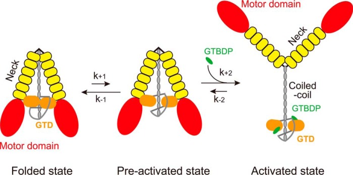
Myosin-5a contains two heavy chains, which are dimerized via the coiled-coil regions. Thus, myosin-5a comprises two heads and two globular tail domains (GTDs). The GTD is the inhibitory domain that binds to the head and inhibits its motor function. Although the two-headed structure is essential for the processive movement of myosin-5a along actin filaments, little is known about the role of GTD dimerization. Here, we investigated the effect of GTD dimerization on its inhibitory activity. We found that the potent inhibitory activity of the GTD is dependent on its dimerization by the preceding coiled-coil regions, indicating synergistic interactions between the two GTDs and the two heads of myosin-5a. Moreover, we found that alanine mutations of the two conserved basic residues at N-terminal extension of the GTD not only weaken the inhibitory activity of the GTD but also enhance the activation of myosin-5a by its cargo-binding protein melanophilin (Mlph). These results are consistent with the GTD forming a head to head dimer, in which the N-terminal extension of the GTD interacts with the Mlph-binding site in the counterpart GTD. The Mlph-binding site at the GTD-GTD interface must be exposed prior to the binding of Mlph. We therefore propose that the inhibited Myo5a is equilibrated between the folded state, in which the Mlph-binding site is buried, and the preactivated state, in which the Mlph-binding site is exposed, and that Mlph is able to bind to the Myo5a in preactivated state and activates its motor function.

Keywords: allosteric regulation; cytoskeleton; intracellular trafficking; molecular motor; myosin; myosin-5a.


Figures

FIGURE 1.
FIGURE 1.
The interactions between the GTD dimer and between Myo5a-GTD and Mlph-GTBDP. A, cartoon representation of the Myo5a-GTD structure (Protein Data Bank code 3WB8) showing a head to tail dimer. There are four putative dimers in the crystal structure of mouse Myo5a-GTD, and only one dimer (chains a and c) is shown here. B, cartoon representation of Myo5b-GTD structure (Protein Data Bank code 4J5M) showing a head to head dimer. C, cartoon representation of the Myo5a-GTD·Mlph-GTBDP complex structure (Protein Data Bank code 4LX2). SD-1 and SD-2 indicate subdomains 1 and 2 of the GTD, respectively. Sticks show the residues at the GTD-GTD interface and the interface between Myo5a-GTD and Mlph-GTBDP. Spheres show the two conserved basic residues Lys1730 and Lys1803, which are essential for inhibiting motor function (7). In the head to tail model of the GTD dimer, the distances between the two Lys1730 and between the two Lys1803 are 79 and 66 Å, respectively. In the head to head model of the GTD dimer, the distances between the two Lys1730 and between the two Lys1803 are 153 and 142 Å, respectively. For clarity, all residue numbers refer to the melanocyte isoform of Myo5a (see “Experimental Procedures” for details).
FIGURE 2.
FIGURE 2.
Shortening of coiled-coil in the Myo5a tail decreases its affinity to Myo5a-HMM. A, diagram of Myo5a structure and Myo5 tail truncation constructs. The coiled-coils were predicted by an online tool (Paircoil). The residue numbers in this study refer to a melanocyte isoform of Myo5a, which is 24 residues longer than that used in most publications. See “Experimental Procedures” for the corresponding residues numbers in most publications. B, SDS-PAGE (4–20%) of purified Myo5a tail proteins. C, the ATPase activity of Myo5a-HMM in the presence of Myo5a tail with different length of coiled-coil. The ATPase activity of Myo5a-HMM was measured in a solution containing 20 mm MOPS-KOH (pH 7.0), 100 mm NaCl, 1 mm MgCl2, 1 mm DTT, 0.25 mg/ml BSA, 12 μm CaM, 0.5 mm ATP, 2.5 mm phosphoenol pyruvate, 20 units/ml pyruvate kinase, 40 μm actin, 1 mm EGTA, 0.03–0.05 μm Myo5a-HMM, and various concentrations of Myo5a tail. The Kd of Myo5a tail to Myo5a-HMM was obtained by a hyperbolic fit. D, the calculated Kd of Myo5a tail to Myo5a-HMM. The Kd values are means ± S.D. from three independent assays of two protein preparations.
FIGURE 3.
FIGURE 3.
Size exclusive chromatography of Myo5a tail constructs. A, Superdex G200 size exclusion chromatography of the purified Myo5a tail constructs. Black squares indicate the elution peaks of each standard proteins. Red triangles indicate the elution peaks of each Myo5a tail constructs. The void volume of the column was 7.7 ml. B, the theoretical and apparent molecular masses of Myo5a tail constructs. The theoretical molecular masses were calculated on the basis of the amino acid composition of Myo5 tail. The apparent molecular masses were estimated from size exclusion chromatography. RKAA, R1490A/K1491A.
FIGURE 4.
FIGURE 4.
Mutation of the conserved basic residues at the GTD-GTD interface of Myo5a GTD dimer decreases the affinity to Myo5a-HMM. A–C, the ATPase activity of Myo5a-HMM was measured in the presence of GST-Myo5a-GTD (A), His-Myo5a-T1344 (B), or His-Myo5a-GTD (C). The ATPase assay was conducted as described in the legend of Fig. 2. D, the Kd of Myo5a tail constructs to Myo5a-HMM. The values are means ± S.D. from three independent assays of two protein preparations.
FIGURE 5.
FIGURE 5.
Effects of R1490A/K1491A mutations on the regulation of Myo5a-FL and the activation by Mlph-GTBDP. A, the ATPase activities of Myo5a-FL under EGTA and pCa4 conditions. The ATPase activity of Myo5a-FL was measured in a solution containing 20 mm MOPS-KOH (pH 7.0), 100 mm KCl, 1 mm MgCl2, 1 mm DTT, 0.25 mg/ml BSA, 12 μm CaM, 0.5 mm ATP, 2.5 mm phosphoenol pyruvate, 20 units/ml pyruvate kinase, 40 μm actin, 1 mm EGTA, and 0.02–0.04 μm Myo5a-FL. EGTA (1 mm) was replaced with 0.9 mm EGTA and 1 mm CaCl2 for pCa4 conditions. The values are means ± S.D. from three independent assays of three protein preparations. B, stimulation of the ATPase activity of Myo5a-FL by Mlph-GTBDP. The ATPase activity was measured in a solution containing 20 mm MOPS-KOH (pH 7.0), 200 mm NaCl, 1 mm MgCl2, 1 mm DTT, 0.25 mg/ml BSA, 12 μm CaM, 0.5 mm ATP, 2.5 mm phosphoenol pyruvate, 20 units/ml pyruvate kinase, 40 μm actin, 1 mm EGTA, 0.03–0.04 μm Myo5a-FL, and various concentrations of the Mlph-GTBDP. Stimulation of the ATPase activity of Myo5a-FL by Mlph-GTBDP was fit to a hyperbola, defining Kd, the apparent dissociation constants of Mlph-GTBDP to Myo5a-FL, which were 32.6 ± 8.2 μm for wild type and 7.3 ± 0.9 μm for R1490A/K1491A. The values are means ± S.D. from three independent assays of three protein preparations. C, GST pulldown of GST-Mlph-GTBDP with FLAG-tagged Myo5a-FL. The pulled down samples and the inputs were analyzed by SDS-PAGE and detected by Western blot using anti-FLAG antibody and anti-GST antibody (top panel). The amounts of pulled down Myo5a-FL were quantified using NIH image program. The bottom panel shows the means ± S.D. of the amount of pulled down Myo5a-FL (relative to WT) from three independent assays of a single protein preparation. D, GST pulldown of GST-Mlph-GTBDP with His-Myo5a-T1235-WT and His-Myo5a-T1344-WT or -RKAA. The pulled down samples were analyzed by SDS-PAGE and detected by Coomassie Blue staining (top panel). The amounts of pulled down His-Myo5a-T1235 and His-Myo5a-T1344 were quantified as described under “Experimental Procedures.” The bottom panel shows the means ± S.D. of the mole of pulled down His-Myo5a-T1344 (relative to His-Myo5a-T1235-WT) from five independent assays of a two protein preparations. RKAA, R1490A/K1491A; IB, immunoblotting.
FIGURE 6.
FIGURE 6.
Proposed mechanism for the activation of Myo5a by Mlph-GTBDP. The inhibited Myo5a molecules are equilibrated between the folded state and the preactivated state. In the folded state, the two GTDs form a head to head dimer via the interaction between the N-terminal extension in one GTD and the Mlph-GTBDP binding site in the counterpart GTD; the GTD dimer interact with the two heads of Myo5a, thus forming a folded, triangular conformation. In the preactivated state, Myo5a also forms a folded, triangular conformation as in the folded state, except that the Mlph-GTBDP binding sites in the GTDs are exposed. Mlph-GTBDP is able to bind to the GTD of Myo5a in preactivated state and allosterically inhibits the binding of GTD to the head, thus inducing the extended conformation and activating the motor function of Myo5a.

References

    1. Cheney R. E., O'Shea M. K., Heuser J. E., Coelho M. V., Wolenski J. S., Espreafico E. M., Forscher P., Larson R. E., and Mooseker M. S. (1993) Brain myosin-V is a two-headed unconventional myosin with motor activity. Cell 75, 13–23 - PubMed
    1. Rodriguez O. C., and Cheney R. E. (2002) Human myosin-Vc is a novel class V myosin expressed in epithelial cells. J. Cell Sci. 115, 991–1004 - PubMed
    1. Zhao L. P., Koslovsky J. S., Reinhard J., Bähler M., Witt A. E., Provance D. W. Jr., and Mercer J. A. (1996) Cloning and characterization of myr 6, an unconventional myosin of the dilute/myosin-V family. Proc. Natl. Acad. Sci. U.S.A. 93, 10826–10831 - PMC - PubMed
    1. Wang F., Thirumurugan K., Stafford W. F., Hammer J. A. 3rd, Knight P. J., and Sellers J. R. (2004) Regulated conformation of myosin V. J. Biol. Chem. 279, 2333–2336 - PubMed
    1. Li X. D., Mabuchi K., Ikebe R., and Ikebe M. (2004) Ca2+-induced activation of ATPase activity of myosin Va is accompanied with a large conformational change. Biochem. Biophys. Res. Commun. 315, 538–545 - PubMed

Publication types

MeSH terms

LinkOut - more resources