. 2016 Jun 10;291(24):12809-12820.
doi: 10.1074/jbc.M116.719377. Epub 2016 Apr 20.

Zinc Finger Homeodomain Factor Zfhx3 Is Essential for Mammary Lactogenic Differentiation by Maintaining Prolactin Signaling Activity

Affiliations

Zinc Finger Homeodomain Factor Zfhx3 Is Essential for Mammary Lactogenic Differentiation by Maintaining Prolactin Signaling Activity

Dan Zhao et al. J Biol Chem. .

Abstract

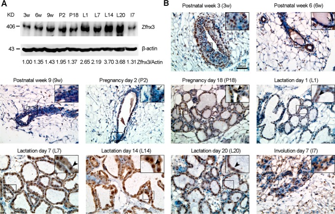
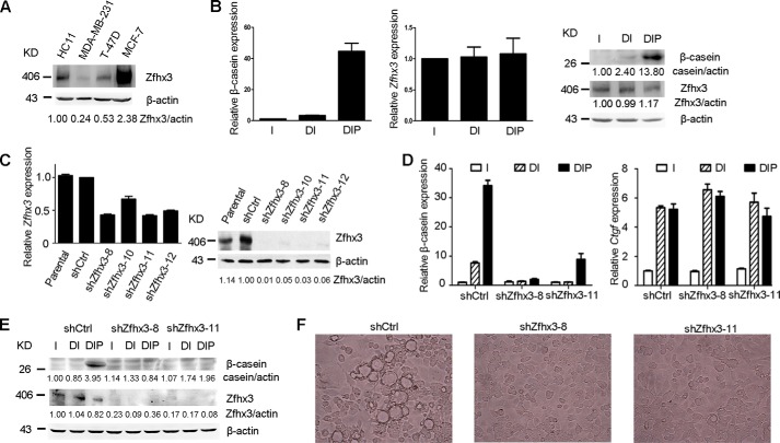
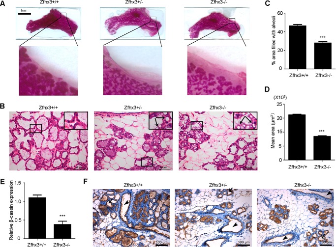
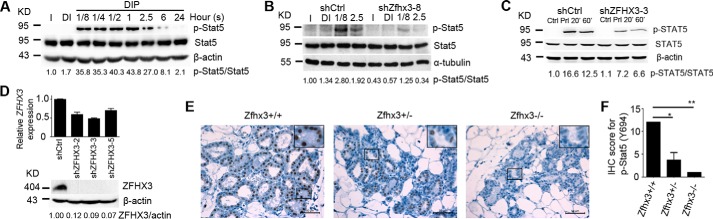
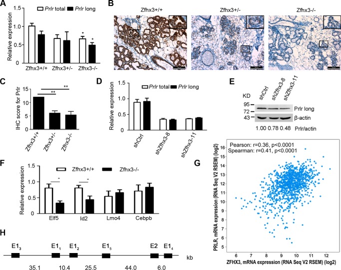
The zinc finger homeobox 3 (ZFHX3, also named ATBF1 for AT motif binding factor 1) is a transcription factor that suppresses prostatic carcinogenesis and induces neuronal differentiation. It also interacts with estrogen receptor α to inhibit cell proliferation and regulate pubertal mammary gland development in mice. In the present study, we examined whether and how Zfhx3 regulates lactogenic differentiation in mouse mammary glands. At different stages of mammary gland development, Zfhx3 protein was expressed at varying levels, with the highest level at lactation. In the HC11 mouse mammary epithelial cell line, an in vitro model of lactogenesis, knockdown of Zfhx3 attenuated prolactin-induced β-casein expression and morphological changes, indicators of lactogenic differentiation. In mouse mammary tissue, knock-out of Zfhx3 interrupted lactogenesis, resulting in underdeveloped glands with much smaller and fewer alveoli, reduced β-casein expression, accumulation of large cytoplasmic lipid droplets in luminal cells after parturition, and failure in lactation. Mechanistically, Zfhx3 maintained the expression of Prlr (prolactin receptor) and Prlr-Jak2-Stat5 signaling activity, whereas knockdown and knock-out of Zfhx3 in HC11 cells and mammary tissues, respectively, decreased Prlr expression, Stat5 phosphorylation, and the expression of Prlr-Jak2-Stat5 target genes. These findings indicate that Zfhx3 plays an essential role in proper lactogenic development in mammary glands, at least in part by maintaining Prlr expression and Prlr-Jak2-Stat5 signaling activity.

Keywords: STAT5 phosphorylation; ZFHX3/ATBF1; development; homeobox; lactogenic differentiation; mammary gland; peptide hormone; prolactin; prolactin receptor (PRLR); transcription factor.


Figures

FIGURE 1.
FIGURE 1.
Dynamic expression of Zfhx3 protein at different stages of postnatal mammary gland development. A, detection of Zfhx3 protein by Western blotting in C57BL/6 female mouse mammary tissues at indicated developmental stages. β-Actin served as the loading control. 3w, postnatal week 3; 6w, postnatal week 6; 9w, postnatal week 9; P2, pregnancy day 2; P18, pregnancy day 18; L1, lactation day 1; L7, lactation day 7; L14, lactation day 14; L20, lactation day 20; I7, involution day 7. The data are representative of three mice per group. Band intensities were quantified using the ImageJ program, and ratios of Zfhx3/β-actin, normalized to that of postnatal week 3, are shown at the bottom. B, IHC staining in mammary glands of C57BL/6 female mice at indicated developmental stages. Arrows show Zfhx3-negative body cells in the terminal end bud (postnatal week 3, prepuberty) or Zfhx3-negative ductal luminal cells at postnatal weeks 6 and 9 (puberty) and pregnancy day 2 (early pregnancy); arrowheads show mature alveolar luminal (Zfhx3-positive) cells in pregnancy day 18 (late pregnancy) and lactation days 1, 7, 14, and 20 (lactation). A representative image from three mice is shown for each stage. Scale bar, 50 μm.
FIGURE 2.
FIGURE 2.
Knockdown of Zfhx3 impairs prolactin-induced differentiation of the mouse HC11 mammary epithelial cells. A, detection of Zfhx3 protein expression by Western blotting in HC11 and three human breast cancer cell lines that show low, moderate, and high expression of ZFHX3, with β-actin as the loading control. B, detection of the expression of β-casein and Zfhx3 in lactogenic hormone-treated HC11 cells through RT-PCR (left and middle panels) and Western blotting (right panel). C, lentiviral expression of shRNAs against Zfhx3 (shZfhx3-8, -10, -11, and -12) knocked down Zfhx3 expression in HC11 cells, as detected by RT-PCR (left panel) and Western blotting (right panel). Parental HC11 cells were used as a positive control. D and E, knockdown of Zfhx3 attenuated prolactin-induced β-casein expression while not affecting the expression of Ctgf, a target gene of dexamethasone but not prolactin, as detected by RT-PCR (D) and Western blotting (E). F, prolactin-induced bubble-like structures, an established indicator of lactogenic hormone-induced differentiation, disappeared upon the knockdown of Zfhx3 in HC11 cells. Cells in B and D–F were treated with insulin (I), insulin plus dexamethasone (DI), or DIP for 5 days; in F only the DIP groups are shown. All Western blots were quantified using the ImageJ program. Ctrl, control.
FIGURE 3.
FIGURE 3.
Deletion of Zfhx3 interferes with the lactogenic development of mouse mammary gland. A and B, underdeveloped mammary glands in Zfhx3 depleted females at lactation day 2, as seen in images of mammary glands from whole mount preparation (A) and H&E staining of tissue sections (B). Arrows in B indicate large cytoplasmic lipid droplets in the luminal epithelial cells of Zfhx3-deleted mammary glands. C, quantification of the total area filled by lactating alveoli in the H&E-stained sections using ImageJ. At least four mice were analyzed, and for each section, at least 20 images (×20) were used to analyze the area occupied by alveoli. D, quantification of the mean area of a single alveoli in the mammary glands using ImageJ. A total of at least 1000 alveoli were analyzed per genotype. For C and D, the data are means ± S.E. E, expression of β-casein by real time PCR; the data are means ± S.E. F, IHC staining of β-casein at lactation in the mammary glands; arrows show empty alveolar lumen, and arrowheads show empty ducts. Statistical significance was calculated by two-tailed Student's t test. ***, p < 0.001.
FIGURE 4.
FIGURE 4.
Zfhx3 is necessary for prolactin-induced phosphorylation of STAT5. A, parental HC11 cells were treated with 5 μg/ml prolactin in a time-dependent manner and Stat5 phosphorylation at tyrosine 694 (p-Y694-Stat5) was detected by Western blotting. B, mouse HC11 mammary epithelial cells with stable knockdown of Zfhx3 (shZfhx3-8) or control (shCtrl) were treated with insulin (I), insulin plus dexamethasone (DI), or DIP for 7.5 min or 2.5 h and then subjected to Western blotting to detect p-Y694-Stat5 and total Stat5. C and D, stable knockdown of ZFHX3 in human breast cancer cell line T-47D also reduced prolactin (Prl)-induced STAT5 phosphorylation, as detected by Western blotting. Knockdown efficiency was confirmed by RT-PCR and Western blotting (D). E and F, IHC staining of p-Y694-Stat5 in lactation day 2 mammary glands (E). IHC score is shown as means ± S.E. (F) and was produced by multiplying the percentage of positive cells (scored as 0 for negative, 1 for < 10%, 2 for 11–50%, 3 for 50–80%, and 4 for > 80%) with the intensity of staining (0 for negative, 1 for weak, 2 for moderate, and 3 for strong). Statistical significance was calculated by one-way analysis of variance followed by Bonferroni's test. *, p < 0.05; **, p < 0.01. All Western blots were quantified using the ImageJ program.
FIGURE 5.
FIGURE 5.
Zfhx3 is needed for the expression of Prlr in mouse mammary epithelial cells. A–C, deletion of Zfhx3 down-regulates Prlr expression, as detected by RT-PCR analysis for Prlr mRNA (total and long form, A) or by IHC staining for Prlr protein (B) at day 2 of lactation. C, IHC score is shown as means ± S.E., statistical significance was calculated by one-way analysis of variance followed by Bonferroni's test; **, p < 0.01). D and E, detection of Prlr expression in HC11 cells with stable knockdown of Zfhx3 by RT-PCR (total and long form, D) or Western blotting (long form, E), band intensities in Western blot were quantified using the ImageJ program. F, Zfhx3-mediated gene regulation in mammary alveolar development. Gene expression analysis of mammary glands from MMTV-Cre/Zfhx3+/+ and MMTV-Cre/Zfhx3f/f mice at day 2 of lactation. At least four mice per genotype were used; the data are means ± S.E. Statistical significance was calculated by two-tailed Student's t test using GraphPad Prism 5; *, p < 0.05. G, the mRNA expression data of ZFHX3 and PRLR in breast invasive carcinoma (TCGA, Provisional) were obtained using cBioPortal. The case set contained 1100 tumor samples. Pearson or Spearman correlation coefficients (r) and two-tailed p value calculations to determine the significance of the correlations were calculated using GraphPad Prism 5. H, the 5′ region of mouse Prlr and the location of five alternative first exons (represented by black boxes). E2, exon 2.

References

    1. Hennighausen L., and Robinson G. W. (2001) Signaling pathways in mammary gland development. Dev. Cell 1, 467–475 - PubMed
    1. Hennighausen L., and Robinson G. W. (2005) Information networks in the mammary gland. Nat. Rev. Mol. Cell Biol. 6, 715–725 - PubMed
    1. Silva C. M., and Shupnik M. A. (2007) Integration of steroid and growth factor pathways in breast cancer: focus on signal transducers and activators of transcription and their potential role in resistance. Mol. Endocrinol. 21, 1499–1512 - PubMed
    1. Brisken C., and O'Malley B. (2010) Hormone action in the mammary gland. Cold Spring Harb. Perspect. Biol. 2, a003178. - PMC - PubMed
    1. Hynes N. E., and Watson C. J. (2010) Mammary gland growth factors: roles in normal development and in cancer. Cold Spring Harb. Perspect. Biol. 2, a003186. - PMC - PubMed

Publication types

MeSH terms

LinkOut - more resources