. 2016 Jun 3;291(23):12014-28.
doi: 10.1074/jbc.M116.721746. Epub 2016 Apr 15.

Global Mapping of O-Glycosylation of Varicella Zoster Virus, Human Cytomegalovirus, and Epstein-Barr Virus

Affiliations

Global Mapping of O-Glycosylation of Varicella Zoster Virus, Human Cytomegalovirus, and Epstein-Barr Virus

Ieva Bagdonaite et al. J Biol Chem. .

Abstract

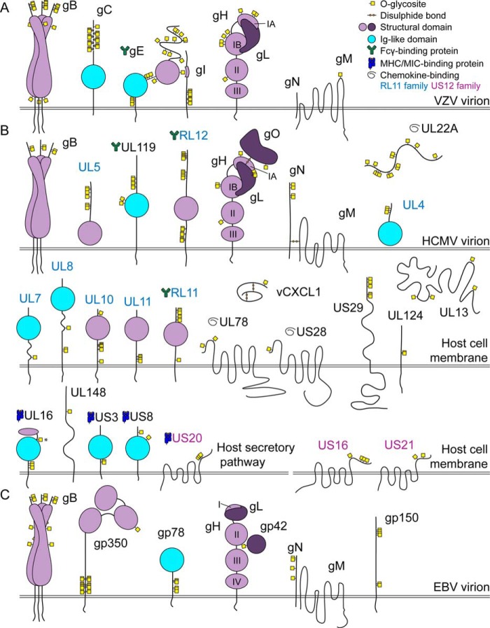
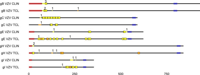
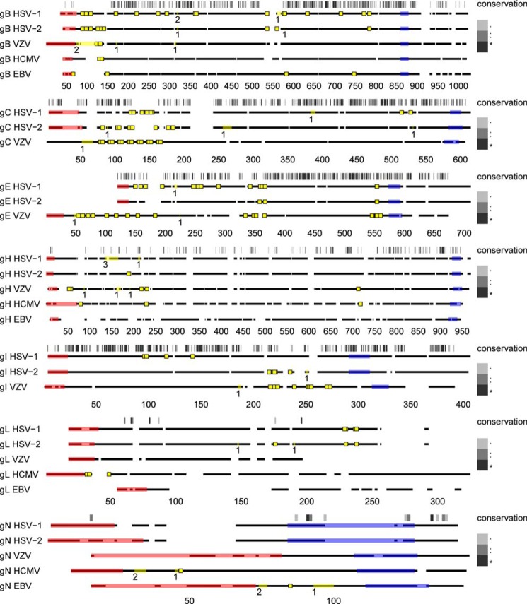
Herpesviruses are among the most complex and widespread viruses, infection and propagation of which depend on envelope proteins. These proteins serve as mediators of cell entry as well as modulators of the immune response and are attractive vaccine targets. Although envelope proteins are known to carry glycans, little is known about the distribution, nature, and functions of these modifications. This is particularly true for O-glycans; thus we have recently developed a "bottom up" mass spectrometry-based technique for mapping O-glycosylation sites on herpes simplex virus type 1. We found wide distribution of O-glycans on herpes simplex virus type 1 glycoproteins and demonstrated that elongated O-glycans were essential for the propagation of the virus. Here, we applied our proteome-wide discovery platform for mapping O-glycosites on representative and clinically significant members of the herpesvirus family: varicella zoster virus, human cytomegalovirus, and Epstein-Barr virus. We identified a large number of O-glycosites distributed on most envelope proteins in all viruses and further demonstrated conserved patterns of O-glycans on distinct homologous proteins. Because glycosylation is highly dependent on the host cell, we tested varicella zoster virus-infected cell lysates and clinically isolated virus and found evidence of consistent O-glycosites. These results present a comprehensive view of herpesvirus O-glycosylation and point to the widespread occurrence of O-glycans in regions of envelope proteins important for virus entry, formation, and recognition by the host immune system. This knowledge enables dissection of specific functional roles of individual glycosites and, moreover, provides a framework for design of glycoprotein vaccines with representative glycosylation.


Figures

FIGURE 1.
FIGURE 1.
The O-glycoproteomes of VZV, HCMV, and EBV. A graphical depiction of identified O-linked glycosylation sites with respect to known structural features of viral glycoproteins (59, 60, 67, 72, 76, 96, 112, 120, 126–128). Domains of gH are labeled with Roman numerals. The O-glycosylation site marked with an asterisk can potentially have a slightly different location because of site ambiguity. Sites mapping to secreted proteins/regions facing the lumenal side of the secretory pathway (>98.5% of glycopeptides) are shown (see all identified glycopeptides in supplemental Data Set S1). A, VZV, all identical tandem repeats are shown occupied in VZV gC. B, HCMV. C, EBV.
FIGURE 2.
FIGURE 2.
O-Glycosites found in VZV derived from infected fibroblasts (TCL) and a clinical sample. VZV (strain Dumas) gB, gC, gE, gH, and gI protein sequences are shown as black lines, drawn to scale. Predicted signal peptides and transmembrane regions are shaded in pink and blue, respectively. Unambiguous O-glycosylation sites are shown as colored squares, whereas ambiguous sites are marked as colored lines within the protein backbone, where the number above indicates the number of glycosites. Trypsin and unique chymotrypsin digestion-derived glycosites are marked in yellow and orange, respectively. All identical potentially glycosylated VZV tandem repeats are shown occupied. Reference VZV sequences were used because of unavailable annotation of investigated isolate sequences. Glycoprotein M is not shown, because it was only found glycosylated in the infected cell lysate. CLIN, clinical sample; TCL, total infected cell lysate.
FIGURE 3.
FIGURE 3.
Conservation of O-linked glycosylation sites on homologous envelope glycoproteins of human herpesviruses. Clustal Omega server was used to align amino acid sequences of gB, gH, gL, and gN between HSV-1 (31), HSV-2 (33), VZV, HCMV, and EBV, as well as gC, gE, and gI between the alphaherpesviruses. Conserved glycoprotein M was not included, because it was only found glycosylated in one of the investigated viruses. Protein backbones are depicted as broken black lines, where spaces represent gaps in the alignment. Individual alignments were drawn to scale (indicated below each graph). Sequence conservation is indicated above the aligned sequences for each set and is represented by a grayscale barcode that maps to the Clustal alignment score, as shown in the legend. In brief, for the Clustal alignment score, an asterisk indicates a position with a fully conserved residue, and a colon indicates conservation of an amino acid with strongly similar properties, whereas a period indicates conservation of an amino acid with weakly similar properties. Predicted signal peptides and transmembrane regions are shaded in pink and blue, respectively. Unambiguous O-glycosylation sites are shown as yellow squares, whereas ambiguous sites are marked as yellow lines within the protein backbone, where the number below indicates the number of glycosites. It should be noted that O-glycosylation sites on VZV are derived both from the clinical sample and the infected total cell lysate. All identical potentially glycosylated VZV tandem repeats are shown occupied. Two ambiguous O-glycosylation sites from our previous publication (31) (HSV-1 gB 109–123 (HexHexNAc) and gE 135–143 (HexHexNAc)) were omitted from the graph, because we cannot exclude the possibility they could be part of an elongated structure on an adjacent site. Reference strain sequences were used for HSV-2, VZV, and EBV because of incomplete or unavailable annotation of investigated strains.

References

    1. Davison A. J. (2002) Evolution of the herpesviruses. Vet. Microbiol. 86, 69–88 - PubMed
    1. Grinde B. (2013) Herpesviruses: latency and reactivation—viral strategies and host response. J. Oral Microbiol. 5, 22766 - PMC - PubMed
    1. McGeoch D. J., Rixon F. J., and Davison A. J. (2006) Topics in herpesvirus genomics and evolution. Virus Res. 117, 90–104 - PubMed
    1. Fishman J. A. (2013) Overview: cytomegalovirus and the herpesviruses in transplantation. Am. J. Transplant. 3, (Suppl. 3) 1–8 - PubMed
    1. Shiley K., and Blumberg E. (2010) Herpes viruses in transplant recipients: HSV, VZV, human herpes viruses, and EBV. Infect. Dis. Clin. North Am. 24, 373–393 - PubMed

Publication types

MeSH terms