. 2016 Jul 1;291(27):13974-13986.
doi: 10.1074/jbc.M116.725978. Epub 2016 Apr 22.

Engineering Highly Potent and Selective Microproteins against Nav1.7 Sodium Channel for Treatment of Pain

Affiliations

Engineering Highly Potent and Selective Microproteins against Nav1.7 Sodium Channel for Treatment of Pain

Anatoly Shcherbatko et al. J Biol Chem. .

Abstract

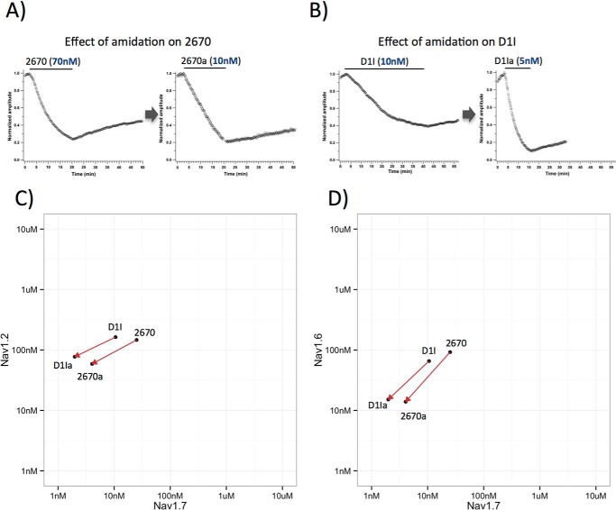
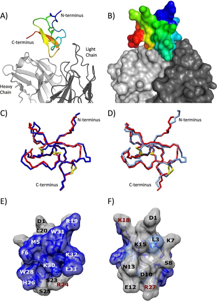
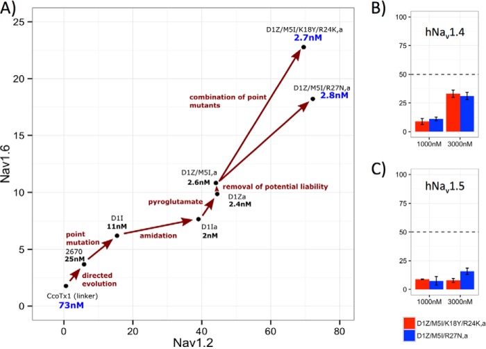
The prominent role of voltage-gated sodium channel 1.7 (Nav1.7) in nociception was revealed by remarkable human clinical and genetic evidence. Development of potent and subtype-selective inhibitors of this ion channel is crucial for obtaining therapeutically useful analgesic compounds. Microproteins isolated from animal venoms have been identified as promising therapeutic leads for ion channels, because they naturally evolved to be potent ion channel blockers. Here, we report the engineering of highly potent and selective inhibitors of the Nav1.7 channel based on tarantula ceratotoxin-1 (CcoTx1). We utilized a combination of directed evolution, saturation mutagenesis, chemical modification, and rational drug design to obtain higher potency and selectivity to the Nav1.7 channel. The resulting microproteins are highly potent (IC50 to Nav1.7 of 2.5 nm) and selective. We achieved 80- and 20-fold selectivity over the closely related Nav1.2 and Nav1.6 channels, respectively, and the IC50 on skeletal (Nav1.4) and cardiac (Nav1.5) sodium channels is above 3000 nm The lead molecules have the potential for future clinical development as novel therapeutics in the treatment of pain.

Keywords: Nav1.7; ceratotoxin; directed evolution; engineering; pain; sodium channel; structure-function; toxin.


Figures

FIGURE 1.
FIGURE 1.
A, amino acid sequence alignment of microproteins included in the initial screening. CcoTx1 and three other molecules were selected for designing the initial libraries on which directed evolution was performed. B, schematic diagram of Nav1.7 with general domain structure. The locations of the four HA tag constructs (M1–M4) that retained a functional Nav1.7 channel are shown. M1 denotes insertion of the GSYPYDVPDYAGS sequence after residue 770, M2 after residue 829, M3 after residue 902, and M4 after residue 1540 (Uniprot Q15858). C, time course of Nav1.7 current block by 250 nm CcoTx1 in HEK293 cells transiently transfected with HA tag-modified Nav1.7 channels. HA tag insertion into the channel extracellular loops S1-S2/D2 (construct M1), S5-S6/D2 (construct M3), or S1-S2/D4 (construct M4) has no influence on CcoTx1 inhibition; however, HA tag insertion into the extracellular loop S3-S4/D2 (construct M2) abolishes the blocking activity of CcoTx1. The manual whole-cell patch clamp technique was used to record Nav1.7 currents. Nav1.7 currents were evoked by a 15-ms step depolarization to 0 mV every 10 s from a holding potential of −90 mV. Data are presented as normalized peak current amplitude versus time. Currents were normalized to the maximum amplitude of control peak current. The solid line indicates the time of compound application. D, amino acid sequence alignment of the S3-S4 region of domain 2 (D2) for several sodium channels (Nav1.1–Nav1.8). Main differences in Nav1.7 are highlighted.
FIGURE 2.
FIGURE 2.
A, potency scatter plot (IC50 Nav1. 7 versus IC50 counter-screening channel Nav1.2) of molecules generated by directed evolution experiments. B, same plot as in A, but with Nav1.6 as counter-screening channel. The starting molecule (CcoTx1) and the molecule selected for saturation mutagenesis and optimization (2670) are depicted in yellow. Increased potency and selectivity are highlighted by black arrows. The three rounds of directed evolution are color-coded black (round 1), blue (round 2), and red (round 3). C–F, certain amino acids that increase selectivity are preferentially found in some positions. Position-specific potency scatter plots are shown (Nav1.7 versus Nav1.2), where each compound's potency on the two channels is represented with a letter corresponding to the amino acid at that specific position. Positions 5, 12, and 20 (C and D) show good separation between amino acids, suggesting clear selectivity preferences. The plots indicate that a methionine achieves better selectivity than tryptophan at position 5; glutamate achieves better selectivity than lysine at position 12, and arginine achieves better selectivity than tyrosine at position 20. In contrast, at position 32 (F), no strong separation is observed between aspartate and lysine.
FIGURE 3.
FIGURE 3.
Results of saturation mutagenesis are expressed as a potency table against Nav1.7 for all single mutant variants screened. Potency (IC50 in nm) is color-coded from red to blue. The color scheme is selected such that the IC50 improvement is red, worsening is blue, and white is no change with respect to initial compound 2670. Light gray shows cysteines that were not mutated in the study. Dark gray shows positions with no data due to low or no expression. Positions with mainly blue cells (such as positions Met-5, Phe-6, Ser-23, Trp-28, Lys-30, and Trp-31) are likely to form the interface with the channel, although positions with mainly white cells (Asp-10, Glu-12, Asn-13, and Lys-15) are likely to be less involved in the interaction. Finally, positions with red cells (Asp-1, Ser-8, and Ser-25) represent mutations that improve the potency of the molecule. The top of the table shows the amino acid sequence of the 2670 microprotein together with the connectivity of the disulfide bridges.
FIGURE 4.
FIGURE 4.
A and B, time course of Nav1.7 current block by the non-amidated and amidated variants of compounds 2670 and D1I in HEK293 cells stably expressing the human Nav1.7 channel. The manual whole-cell patch clamp technique was used to record Nav1.7 currents. Nav1.7 currents were evoked by a 15-ms step depolarization to 0 mV every 10 s from a holding potential of −90 mV. Currents were normalized to the maximum amplitude of control peak current. Data presented as normalized peak current amplitude versus time. The time of compound application is indicated by solid line. C and D, effect of amidation for 2670 and D1I illustrated in selectivity plots. Amidation increases potency for both compounds on Nav1.7 but also on Nav1.2 and Nav1.6. Improvement in potency is accompanied by selectivity benefits against Nav1.2 but not Nav1.6.
FIGURE 5.
FIGURE 5.
A, ribbon representation of the crystal structure of variant 2670 in complex with 6F1 Fab fragment. 2670 is colored in rainbow gradient going from N terminus (blue) to C terminus (red). 6F1 Fab fragment is shown in gray, with only part of the structure visible. B, surface representation of the complex in the same orientation and color-coding as in A. C, backbone representation of variant 2670 (red) superposed to Huwentoxin-IV (blue) resulting in an r.m.s.d. of 0.9 Å. D, backbone representation of variant 2670 (red) superposed to Hainantoxin-IV (light blue) resulting in an r.m.s.d. of 0.6Å. Cysteine disulfide bridges are shown in stick representation in both C and D. E and F, functional epitope (blue and cyan) of 2670 against Nav1.7 channel mapped on crystal structure of 2670. Functional epitope is defined as all positions for which the fraction of substitutions worsening potency by a factor 2 or more is larger than a threshold (blue, at least 75%; cyan, 50–75%).
FIGURE 6.
FIGURE 6.
A, evolution of compounds starting from CcoTx1 up to the rationally engineered multiple mutants (top-right corner). Selectivity to Nav1.2 and Nav1.6 is reported on the axis and potency is illustrated in nm on the plot. Effect of D1Z/M5I/K18Y/R24K,a and D1Z/M5I/R27N,a on human (B) Nav1.4 and (C) Nav1.5. The microprotein activity was tested at a concentration of 1000 and 3000 nm and percent inhibition was measured. Even at 3000 nm neither microprotein reached 50% inhibition on either channel, giving better than 1000-fold selectivity (hNav1.7 potency of these microproteins is ∼2.7 nm; A).

References

    1. Goldberg D. S., and McGee S. J. (2011) Pain as a global public health priority. BMC Public Health 11, 770. - PMC - PubMed
    1. Cox J. J., Reimann F., Nicholas A. K., Thornton G., Roberts E., Springell K., Karbani G., Jafri H., Mannan J., Raashid Y., Al-Gazali L., Hamamy H., Valente E. M., Gorman S., Williams R., et al. (2006) An SCN9A channelopathy causes congenital inability to experience pain. Nature 444, 894–898 - PMC - PubMed
    1. Goldberg Y. P., MacFarlane J., MacDonald M. L., Thompson J., Dube M. P., Mattice M., Fraser R., Young C., Hossain S., Pape T., Payne B., Radomski C., Donaldson G., Ives E., Cox J., et al. (2007) Loss-of-function mutations in the Nav1.7 gene underlie congenital indifference to pain in multiple human populations. Clin. Genet. 71, 311–319 - PubMed
    1. Yang Y., Wang Y., Li S., Xu Z., Li H., Ma L., Fan J., Bu D., Liu B., Fan Z., Wu G., Jin J., Ding B., Zhu X., and Shen Y. (2004) Mutations in SCN9A, encoding a sodium channel alpha subunit, in patients with primary erythermalgia. J. Med. Genet. 41, 171–174 - PMC - PubMed
    1. Fertleman C. R., Baker M. D., Parker K. A., Moffatt S., Elmslie F. V., Abrahamsen B., Ostman J., Klugbauer N., Wood J. N., Gardiner R. M., and Rees M. (2006) SCN9A mutations in paroxysmal extreme pain disorder: allelic variants underlie distinct channel defects and phenotypes. Neuron 52, 767–774 - PubMed

MeSH terms

Substances