. 2016 Jun 3;36(3):e00337.
doi: 10.1042/BSR20150278. Print 2016 Jul.

Expression signature of lncRNAs and their potential roles in cardiac fibrosis of post-infarct mice

Affiliations

Expression signature of lncRNAs and their potential roles in cardiac fibrosis of post-infarct mice

Xuefeng Qu et al. Biosci Rep. .

Abstract

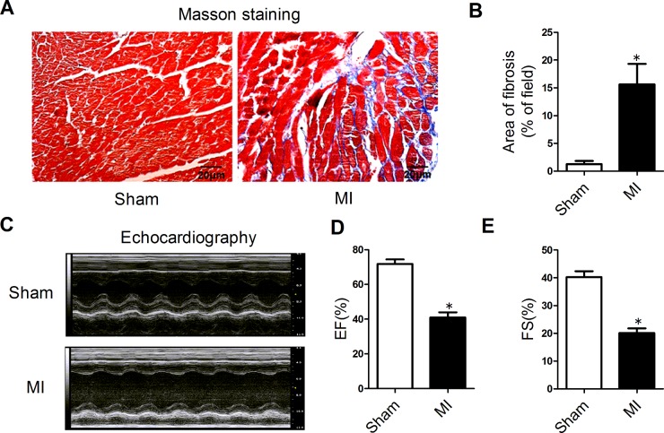
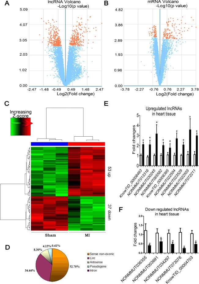
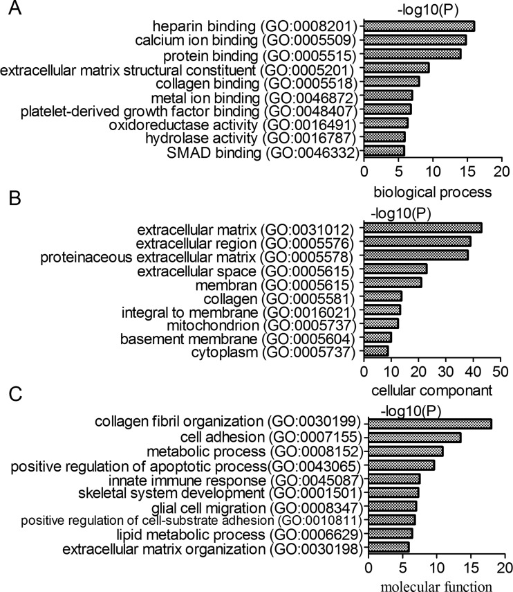
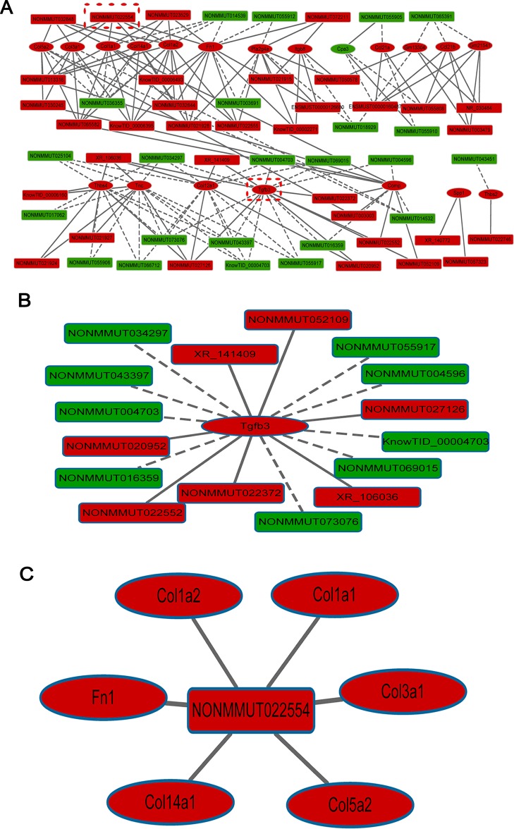
The present study aimed to investigate whether long non-coding RNAs (lncRNAs) are involved in cardiac fibrogenesis induced by myocardial infarction (MI). The differentially expressed lncRNAs and mRNAs in peri-infarct region of mice 4 weeks after MI were selected for bioinformatic analysis including gene ontology (GO) enrichment, pathway and network analysis. Left ventricular tissue levels of lncRNAs and mRNAs were compared between MI and sham control mice, using a false discovery rate (FDR) of <5%. Out of 55000 lncRNAs detected, 263 were significantly up-regulated and 282 down-regulated. Out of 23000 mRNAs detected, 142 were significantly up-regulated and 67 down-regulated. Among the differentially expressed lncRNAs, 53 were up-regulated by ≥2.0-fold change and 37 down-regulated by ≤0.5-fold change. Nine up-regulated and five down-regulated lncRNAs were randomly selected for quantitative real-time PCR (qRT-PCR) verification. GO and pathway analyses revealed 173 correlated lncRNA-mRNA pairs for 57 differentially expressed lncRNAs and 20 differentially expressed genes which are related to the development of cardiac fibrosis. We identified TGF-β3 as the top-ranked gene, a critical component of the transforming growth factor-β (TGF-β) and mitogen activated protein kinase (MAPK) signalling pathways in cardiac fibrosis. NONMMUT022554 was identified as the top-ranked lncRNA, positively correlated with six up-regulated genes, which are involved in the extracellular matrix (ECM)-receptor interactions and the phosphoinositid-3 kinase/protein kinase B (PI3K-Akt) signalling pathway. Our study has identified the expression signature of lncRNAs in cardiac fibrosis induced by MI and unravelled the possible involvement of the deregulated lncRNAs in cardiac fibrosis and the associated pathological processes.

Keywords: cardiac fibrosis; expression; gene; long non-coding RNA (lncRNA); myocardial infarction.


Figures

Figure 1
Figure 1. Cardiac fibrosis induced by myocardial infarction
(A) Representative sections of heart with Masson's trichrome staining at a magnification of 400×. The fibrotic tissues were stained blue. (B) Collagen deposition was quantified using an automated image analyser and is expressed as the percentage of tissue area (n=4). (C) Representative M-mode echocardiography of mice 4 weeks after MI. (D) EF in the MI mice and sham-operated animals. (E) FS in the MI and sham groups (n=6). Data are expressed as mean ± S.E.M.; *P<0.05 compared with sham.
Figure 2
Figure 2. Differentially expressed lncRNAs and mRNAs between MI mice and sham control animals
Volcano plot provides a convenient way to visualize the distribution of datasets of lncRNAs (A) and mRNAs (B). (C) The results from hierarchical clustering showing the distinct lncRNA expression profiles between MI and control. ‘Red’ indicates high relative expression and ‘green’ low relative expression. (D) Distribution of differentially expressed lncRNAs according to different biotypes. (E and F) Verification of the microarray results using qRT-PCR methods. Nine up-regulated and five down-regulated lncRNAs from the infracted hearts were studied using β-actin as an internal control (n=5–6). *P<0.05 compared with sham-operated control.
Figure 3
Figure 3. Gene ontology enrichment analysis of differentially expressed mRNAs
(A) Biological process enrichment analysis. (B) Cell component enrichment analysis. (C) Molecular function enrichment analysis.
Figure 4
Figure 4. Prediction of lncRNA–mRNA association network
(A) The co-expression network was composed of 173 connections between 57 lncRNAs and 20 coding genes. (B) Connections between top-ranked gene TGF-β3 and lncRNAs. (C) Connections between the top-ranked lncRNA NONMMUT022554 and coding genes. Rectangle nodes represent mRNAs and hexagon nodes represent lncRNAs. Red nodes represent up-regulated lncRNAs or mRNAs and green nodes represent down-regulated lncRNAs or mRNAs. The solid line indicates positive correlation and the dashed line negative correlation.

References

    1. Leask A. Getting to the heart of the matter: new insights into cardiac fibrosis. Circ. Res. 2015;116:1269–1276. doi: 10.1161/CIRCRESAHA.116.305381. - DOI - PubMed
    1. Euler G. Good and bad sides of TGFbeta-signaling in myocardial infarction. Front. Physiol. 2015;6:66. doi: 10.3389/fphys.2015.00066. - DOI - PMC - PubMed
    1. Hafizi S., Wharton J., Chester A.H., Yacoub M.H. Profibrotic effects of endothelin-1 via the ETA receptor in cultured human cardiac fibroblasts. Cell Physiol. Biochem. 2004;14:285–292. doi: 10.1159/000080338. - DOI - PubMed
    1. Siddesha J.M., Valente A.J., Sakamuri S.S., Yoshida T., Gardner J.D., Somanna N., Takahashi C., Noda M., Chandrasekar B. Angiotensin II stimulates cardiac fibroblast migration via the differential regulation of matrixins and RECK. J. Mol. Cell. Cardiol. 2013;65:9–18. doi: 10.1016/j.yjmcc.2013.09.015. - DOI - PMC - PubMed
    1. Ponten A., Folestad E.B., Pietras K., Eriksson U. Platelet-derived growth factor D induces cardiac fibrosis and proliferation of vascular smooth muscle cells in heart-specific transgenic mice. Circ. Res. 2005;97:1036–1045. doi: 10.1161/01.RES.0000190590.31545.d4. - DOI - PubMed

Publication types

MeSH terms

Substances