. 2017 Feb 1;81(3):231-242.
doi: 10.1016/j.biopsych.2016.02.013. Epub 2016 Feb 21.

Neurochemical Mediation of Affiliation and Aggression Associated With Pair-Bonding

Affiliations

Neurochemical Mediation of Affiliation and Aggression Associated With Pair-Bonding

Kyle L Gobrogge et al. Biol Psychiatry. .

Abstract

Background: The neuropeptides vasopressin and corticotropin-releasing factor facilitate, while serotonin inhibits, aggression. How the brain is wired to coordinate interactions between these functionally opposed neurotransmitters to control behavioral states is poorly understood.

Methods: Pair-bonded male prairie voles (Microtus ochrogaster) were infused with a retrograde tracer, Fluoro-Gold, and tested for affiliation and aggression toward a female partner or novel female subject. Subsequent immunocytochemical experiments examined neuronal activation using Fos and neurochemical/neuroreceptor profiles on brain areas involved in these social behaviors. Finally, a series of behavioral pharmacologic and real-time in vivo brain microdialysis experiments were performed on male prairie voles displaying affiliation or aggression.

Results: We localized a subpopulation of excitatory vasopressin neurons in the anterior hypothalamus that may gate corticotropin-releasing factor output from the amygdala to the anterior hypothalamus and then the lateral septum to modulate aggression associated with mate guarding. Conversely, we identified a subset of inhibitory serotonergic projection neurons in the dorsal raphe that project to the anterior hypothalamus and may mediate the spatiotemporal release of neuropeptides and their interactions in modulating aggression and affiliation.

Conclusions: Together, this study establishes the medial extended amygdala as a major neural substrate regulating the switch between positive and negative affective states, wherein several neurochemicals converge and interact to coordinate divergent social behaviors.

Keywords: Anterior hypothalamus; Corticotropin-releasing factor; Dopamine; Medial amygdala; Serotonin; Vasopressin.


Conflict of interest statement

All authors report no biomedical financial interests or potential conflicts of interest.

Figures
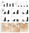
Figure 1
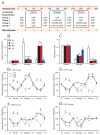
Figure 1. Differential Limbic Circuit Activation Associated with Affiliation and Aggression
(A) Male prairie voles that pair-bonded with a female for two weeks displayed robust aggression against a novel female but (B) high levels of affiliation toward their familiar female partner. This behavioral pattern was consistent across animals that received FG injections into the lateral septum (LS), nucleus accumbens (NAcc), anterior hypothalamic nucleus (AH) or posterior dorsal medial amygdala (MeAPD) with significant main (F(2,4,20) = 32.58, p < 0.001 for aggression and F(2,4,20) = 37.29, p < 0.001 for affiliation), but not interaction (F(2,4,20) = 1.37, ns, for aggression and F(2,4,20) = 1.79, ns, for affiliation), effects. In these four FG injection groups, aggression against a novel female induced a significant increase in the density of FOS-ir neurons in the AH (C & D) and MeAPD (C–E) while affiliation induced a significant increase in the density of FOS-ir neurons in the DR compared to controls with significant main (F(2,4,20) = 6.85, p < 0.05 for aggression and F(2,4,20) = 7.64, p < 0.05 for affiliation), but not interaction (F(2,4,20) = 1.13, ns, for aggression and F(2,4,20) = 1.32, ns, for affiliation), effects. In addition, aggression induced a significant increase in the density of neurons double-labeled for FOS-ir/FG-ir in the AH (F) and MeAPD (H) compared to affiliation or controls with significant main (F(2,4,20) = 5.93, p < 0.05), but not interaction (F(2,4,20) = 1.68, ns), effects. Conversely, males displaying affiliation toward their female partner had an increased density of FOS-ir/FG-ir double-labeling in the dorsal raphe nucleus (DR) compared to males displaying aggression against a novel female and controls with significant main (F(2,4,20) = 5.43, p < 0.05), but not interaction (F(2,4,20) = 1.09, ns), effects (H). Light-field photomicrographs (30μm stack) of neurons labeled for FG-ir (brown cytoplasmic staining), FOS-ir (black nuclear staining) or both in the AH (FG injected into LS; I), MeAPD (FG injected into AH; J), and DR (FG injected into AH; K) of males exposed to a stranger female. The open black circles shown in panels I, J, and K depicts the area at higher magnification (5μm stack) in the inset. F: fornix; OT: optic tract; CA: cerebral aqueduct. Bars indicate means ± standard error of the mean. Asterisks (*) indicate, **: p < 0.01 (A & B). Bars labeled with different letters (C–H) differ significantly by post hoc Student Newman-Keuls (SNK) tests of significance examining both main effects and interactions with analysis of variance p value set to < .05. Scale bar = 100μm. The insert within each panel shows neurons double-labeled for FOS-ir/FG-ir, while scale bar = 10μm. See also Tables S1.
Figure 2
Figure 2. AH-5-HT-1a-type Receptor Activation Attenuates AVP- and Pair-Bonding-Induced Aggression
(A) AVP, (B) 5-HT, (C) 5-HTr1a, and (D) AVP/5-HT/5-HTr1a labeling in the anterior hypothalamic nucleus. AVP/5-HTr1a double-labeled neurons indicated by white arrowheads. For the behavioral experiments (E–H), sexually naïve male prairie voles received intra-AH injections of AVP (500 ng in 200 nl CSF)/side) or AVP with a low (0.5 μg/side) or high (5 μg/side) dose of a 5-HTr1a agonist (R(+)-8-OH-DPAT) followed by a 10-min resident intruder test (RIT) toward a novel female. Both doses of the 5-HTr1a agonist blocked AH-AVP-induced aggression (F(2,18) = 9.23, p < 0.01) (E) and enhanced affiliation (F(2,18) = 4.20, p < 0.05) (F) compared to CSF-injected controls. Male prairie voles that were pair-bonded with a female for two weeks were divided into three groups that received intra-AH infusions of CSF (200 nl/side) or CSF containing a 5-HTr1a agonist (5 μg/side) or antagonist (p-MPPI; 5 μg/side) followed by a 10-min RIT. (G & H) Males treated with the 5-HTr1a agonist displayed a significant decrease in aggression (F (2,20) = 40.83, p < 0.001) and an increase in affiliation (F(2,20) = 8.01, p < 0.01) compared to CSF-injected controls. (G) In contrast, males treated with the 5-HTr1a antagonist displayed enhanced aggression toward a novel female than did CSF-injected controls (F(2,20) = 40.83, p < 0.001). Lastly, we tested the behavioral consequences of AVP administration in the AH of pair-bonded males treated with a combination of 5-HTr1a agonist/antagonist infusions. Overall, males displayed significantly higher levels of offensive aggression and low affiliation toward a sexually-receptive female (I & J). Males treated with the 5-HTr1a 5μg antagonist exhibited significantly higher levels of offensive aggression (F (2,20) = 5.39, p < 0.05; I). The effect of enhancing aggressive responding by blocking AH-5-HTr1a, in intra-AH-AVP infused sexually naïve males (E–H), is abolished in pair-boned - intra-AH-AVP treated males - while 5-HTr1a antagonist treatment in these males enhances offensive aggression (I). Thus, the effect of 5-HT1aR activation on aggression can be blocked by AVP, whereas the effect of AVP cannot be blocked by 5-HT antagonists. Bars indicate means ± standard error of the mean. Bars with different alphabetical letters differ significantly from each other. Scale bar = 10μm.
Figure 3
Figure 3. 5-HT-, CRH-, and AVP-Expressing Neurons/Receptors Co-localize in the AH
Photomicrographs displaying cytochemical marker fluorescence histochemistry in the anterior hypothalamic nucleus. (A) serotonin (5-HT) 1a-type receptors (5-HTr1a), (B) vasopressin (AVP), and (C) 5-HTr1a/AVP double-labeled neurons indicated by white arrowheads. (D) 5-HTr1a receptors, (E) corticotrophin-releasing hormone (CRH), and (F) 5-HTr1a/CRH double-labeled neurons indicated by white arrowheads. (G) CRH-type-2 receptors (CRHR2), (H) AVP, and (I) CRHR2/AVP double-labeled neurons indicated by white arrowheads. (J) AVP-V1a-type receptors (V1aR), (K) CRH, (L) Fluorogold (FG - injected into the lateral septum), (M) V1aR/CRH/FG triple-labeled neurons indicated by white arrows. Scale bar = 10μm.
Figure 4
Figure 4. Behaviorally- and Pharmacologically-Evoked Neurotransmitter Release in the AH Reveals Dynamic Regulation of Behavioral Switches
(A) Real-time in-vivo brain microdialysis paradigm. Pair-bonded males were stereotaxically implanted with a microdialysis probe aimed at the anterior hypothalamic nucleus (AH) and divided into four pharmacological treatment groups for vehicle (CSF) infusions with manipulations of vasopressin (AVP), corticotrophin-releasing hormone (CRH), and serotonin (5-HT) systems, respectively. Microdialysate samples were collected every 30-minutes over a 5-hour period in which males were reunited with their female partner, introduced to a novel female, and then re-exposed to their female partner again. Reverse dialysis of pharmacological compounds were infused during exposure to novel females and re-exposure to their female partner. (B & C) Blockade of AVP-V1a-type receptors (V1aRs) or CRH type-2 receptors (CRHR2s) or activation of 5-HT-1a-type receptors (5-HTr1as) in the AH abolished aggression (F(3,28) = 56.25, p < 0.001; B) and facilitated affiliation (F(3,28) = 41.61, p < 0.001; C) toward novel females. Activation of either V1aR or CRHR2 induced aggression (F(3,28) = 18.26, p < 0.01; B) and reduced affiliation (F(3,28) = 15.92, p < 0.01; C) toward their female partner, while blockade of 5-HTr1a decreased affiliation (F(3,28) = 63.74, p < 0.001; C) but did not induce aggression toward their female partner (F(3,28) = 2.91, ns; B). (D) In CSF control males, 5-HT release increased while AVP/CRH release decreased when males were either reunited or re-exposed (F(3,28) = 14.52, p < 0.01) to their female partner. A reverse pattern of neurotransmitter release was found when males were fighting novel females (F(3,28) = 17.49, p < 0.01). (E) Blockade of V1aR in the AH attenuated AVP/CRH release associated with exposure to novel females (F(3,28) = 13.46, p < 0.01) while activation of V1aR facilitated AVP/CRH release and decreased 5-HT release in the AH during partner re-exposure (F(3,28) = 15.82, p < 0.01). (F) Blockade of CRHR2 also diminished AVP/CRH release associated with exposure to novel females (F(3,28) = 14.88, p < 0.01) while activation of CRHR2 enhanced AVP/CRH release and decreased 5-HT release during partner re-exposure (F(3,28) = 13.37, p < 0.01). (G) Activation of 5-HTr1a attenuated AVP/CRH release associated with exposure to novel females (F(3,28) = 18.59, p < 0.01) while blockade of 5-HTr1a diminished increased 5-HT release and decreased AVP/CRH release during partner re-exposure. Bars indicate means ± standard error of the mean. Bars with different alphabetical letters differ significantly from each other at p < 0.01. Line time points indicate percent change from baseline ± standard error of the mean. *: p < 0.01. ANT: antagonist, AGO: agonist, B: baseline (with CSF infusions). See also Figure S1D.
Figure 5
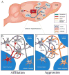
Figure 5. Microcircuit “Switch” Mechanism Programming Behavioral State
(A) Schematic illustrates the neurocircuitry and neurotransmitter circuit phenotypes summarized from monosynaptic neuronal tract-tracing and histochemical experiments. Black arrows indicate anatomical connections and neurochemical projections are color-coded. The anterior hypothalamic nucleus (AH) projects to forebrain areas: ventral pallidum (VP) and bed nucleus of the stria terminalis (BNST) that are involved in pair-bonding behavior; intersects several dopaminergic regions including the ventral tegmental area (VTA), nucleus accumbens (NAcc), caudate putamen (CP), and prefrontal cortex (PFC); integrates olfactory and pheromonal information from the vomeronasal organ through the accessory olfactory bulb (AOB) via the posterior dorsal medial amygdala (MeAPD); receives corticotrophin-releasing hormone (CRH) projections from the MeAPD and serotonergic (5-HT-ergic) input from the dorsal raphe nucleus (DR); and sends CRH projections to the lateral septum (LS). (B) During affiliation, a subset of 5-HT neurons in the DR project axonal collaterals and release 5-HT in the AH. Released 5-HT acts on post-synaptic 5-HT1a-type receptors (5-HTr1a) co-expressed on AVP- and CRH-containing interneurons, in the medial supra-optic nucleus (mSON) and nucleus circularis (NC), to suppress local AVP/CRH release, enhance affiliation, and inhibit aggression. (C) During aggression, a subpopulation of CRH neurons in the MeAPD project dendritic arbors to and release CRH in the AH. Released CRH binds to post-synaptic CRH-type-2 receptors (CRHR2s) co-expressed on the membrane surface of AVP interneurons, in the AH, to facilitate local AVP release. Released AVP then acts on post-synaptic V1a-type receptors (V1aR) co-expressed on a subset of CRH neurons projecting to the LS, where CRH is released and activates CRHR2-expressing neurons to escalate aggression.

Comment in

Similar articles

Cited by

References

    1. Yu Q, Teixeira CM, Mahadevia D, Huang Y, Balsam D, Mann JJ, et al. Dopamine and serotonin signaling during two sensitive developmental periods differentially impact adult aggressive and affective behaviors in mice. Mol Psychiatry. 2014;19(6):688–98. - PMC - PubMed
    1. Miczek KA, Takahashi A, Gobrogge KL, Hwa LS, de Almeida RM. Escalated Aggression in Animal Models: Shedding New Light on Mesocorticolimbic Circuits. Curr Opin Behav Sci. 2015;3:90–5. - PMC - PubMed
    1. Motta SC, Goto M, Gouveia FV, Baldo MV, Canteras NS, Swanson LW. Dissecting the brain’s fear system reveals the hypothalamus is critical for responding in subordinate conspecific intruders. Proc Natl Acad Sci U S A. 2009;106(12):4870–5. - PMC - PubMed
    1. Luiten PG, Koolhaas JM, de Boer S, Koopmans SJ. The cortico-medial amygdala in the central nervous system organization of agonistic behavior. Brain Res. 1985;332(2):283–97. - PubMed
    1. Choi GB, Dong HW, Murphy AJ, Valenzuela DM, Yancopoulos GD, Swanson LW, et al. Lhx6 delineates a pathway mediating innate reproductive behaviors from the amygdala to the hypothalamus. Neuron. 2005;46(4):647–60. - PubMed

Publication types

MeSH terms

LinkOut - more resources