. 2016 Jun 24;291(26):13730-42.
doi: 10.1074/jbc.M116.733766. Epub 2016 Apr 29.

Secreted Frizzled-related Protein 2 (sFRP2) Redirects Non-canonical Wnt Signaling from Fz7 to Ror2 during Vertebrate Gastrulation

Affiliations

Secreted Frizzled-related Protein 2 (sFRP2) Redirects Non-canonical Wnt Signaling from Fz7 to Ror2 during Vertebrate Gastrulation

Eva-Maria Brinkmann et al. J Biol Chem. .

Abstract

Convergent extension movements during vertebrate gastrulation require a balanced activity of non-canonical Wnt signaling pathways, but the factors regulating this interplay on the molecular level are poorly characterized. Here we show that sFRP2, a member of the secreted frizzled-related protein (sFRP) family, is required for morphogenesis and papc expression during Xenopus gastrulation. We further provide evidence that sFRP2 redirects non-canonical Wnt signaling from Frizzled 7 (Fz7) to the receptor tyrosine kinase-like orphan receptor 2 (Ror2). During this process, sFRP2 promotes Ror2 signal transduction by stabilizing Wnt5a-Ror2 complexes at the membrane, whereas it inhibits Fz7 signaling, probably by blocking Fz7 receptor endocytosis. The cysteine-rich domain of sFRP2 is sufficient for Ror2 activation, and related sFRPs can substitute for this function. Notably, direct interaction of the two receptors via their cysteine-rich domains also promotes Ror2-mediated papc expression but inhibits Fz7 signaling. We propose that sFRPs can act as a molecular switch, channeling the signal input for different non-canonical Wnt pathways during vertebrate gastrulation.

Keywords: Fz7; Ror2; Wnt signaling; Xenopus; development; embryo; papc; sFRP2; zebrafish.


Figures

FIGURE 1.
FIGURE 1.
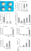
Gain and loss of sFRP2 impairs CE movements in Xenopus. A, representative phenotypes of embryos injected into the dorsal equatorial zone at the four-cell stage with the indicated Mo (15 ng) and synthetic mRNA (500 pg of sfrp2/hsfrp2 or 200 pg for the Mo rescue). B, quantification of the phenotypic analysis shown in A. The numbers of analyzed embryos are indicated on top of the columns. C, for the AC elongation assay, embryos were injected at the two-cell stage with the indicated synthetic mRNAs (300 pg) and excised at stage 9. ACs were cultured with or without Activin overnight to analyze elongation. D, analysis of xbra expression of 10 ACs as shown in C, harvested after 2-h incubation in Activin. Shown is a representative with technical triplicates. This was confirmed by at least two independent experiments with similar results using sibling animal caps of those shown in C and E. E, quantification of the AC elongation shown in C and categorized as not elongated (blue), partially elongated (white), and fully elongated (gray) ACs. The numbers of analyzed ACs are indicated on top of the columns. F, AC elongation assay of embryos injected with 100 pg of each indicated synthetic mRNA. To balance injected mRNA quantities, the respective amount of gfp mRNA was co-injected. Scoring and quantification was as in C and E. Co, control.
FIGURE 2.
FIGURE 2.
sFRP2 is required for papc expression and enhances Ror2-mediated signaling. A, representative papc expression pattern analyzed by whole mount in situ hybridization of gastrula (stage 10.5) embryos injected at the four-cell stage in the dorsal equatorial region with the indicated Mo (15 ng). Arrowheads indicate the site of injection. Co, control. B, quantification of in situ hybridization shown in A. The numbers of analyzed embryos are indicated on top of the columns. C and D, relative (rel) expression of papc analyzed by qPCR in whole embryos injected with the indicated Mos (15 ng) and synthetic mRNA (200 pg). E and F, relative expression of papc analyzed by qPCR in AC explants injected with the indicated synthetic mRNAs (500 pg of ror2/sfrp2 and 150 pg of wnt5a). G, relative expression of ror2 in ACs from uninjected embryos cut at stage 9 and harvested after 14 h with or without Activin (50 ng/ml) when sibling embryos reached stage 25. H, ATF luciferase reporter assay of stage 12 gastrula embryos injected with the indicated synthetic mRNAs (500 pg of ror2/sfrp2 and 100 pg of wnt5a) and the ATF2 firefly and pRL-TK Renilla luciferase reporter constructs. C and G show the mean ± S.D. of three independent experiments. *, p < 0.05 compared with controls. D–F show a representative with technical triplicates. This was confirmed by at least two (D and F) or three (E) independent experiments in different batches of X. laevis with similar results. H shows the mean ± S.D. of biological triplicates of pools of seven embryos each (*, p < 0.05 compared with controls). This was confirmed in at least three independent experiments in different batches of X. laevis.
FIGURE 3.
FIGURE 3.
sFRP2 interacts with Ror2 in HEK293T cells and stabilizes Wnt5a-Ror2 complexes. A, co-IP in HEK293T cells transfected with Ror2-myc- and HA-tagged sFRP2 (1 μg of each). Protein lysates were precipitated with a mouse antibody against myc or HA, respectively, and IgG as a negative control. Western blotting (WB) analysis with anti-HA antibody shows that sFRP2 is pulled down together with Ror2. B, IP from conditioned media generated in HEK293T cells transfected with either Ror2 ECD-FLAG or sFRP2-HA. Purified proteins were mixed and further precipitated with a mouse anti-HA antibody. WB analysis with rabbit anti-FLAG shows that purified sFRP2 binds to purified Ror2-ECD. As a negative control (Ctrl), Ror2 ECD-FLAG conditioned medium was precipitated with anti-HA-coupled magnetic beads and sFRP2-HA conditioned medium with anti-FLAG-coupled magnetic beads. C, the Wnt5a-V5 binding assay was performed in HEK293T cells transfected with Ror2 ECD-FLAG alone or co-transfected with sFRP2-HA, CRD-HA, DKK3-HA, or Fz7-myc (1 μg of each). Each cell sample was treated with equal amounts of Wnt5a-V5 conditioned medium for 15 min before cells were lysed for rabbit anti-FLAG pulldown to precipitate Ror2. The samples were then analyzed on two different WBs. The WB on top was analyzed with anti-V5 antibody for the Wnt5a-V5 fraction bound to precipitated Ror2-FLAG. The bottom WB was analyzed with mouse anti-FLAG and shows that equal amounts of Ror2 were precipitated in the different samples. D, the WB analyzed with mouse anti-HA/anti-myc antibody shows that all co-transfected proteins were expressed (input, first four lanes) and were co-precipitated with the Ror2-FLAG pulldown (IP, α-FLAG, last four lanes). Only Dkk3 was not precipitated with Ror2. A schematic of the different proteins used in the experiment is shown below. sFRP family proteins are related to Fz receptors in the CRD. TM, transmembrane domain; CD, cytoplasmic domain; Cys, cysteine-rich domain. E, confocal microscopy analysis of live zebrafish embryos expressing 1 ng of mRNA of the indicated constructs at 30–50% epiboly stages shown in the indicated colors. Confocal images represent single z sections. Ror2-mCherry shows membrane localization regardless of the presence of Wnt5a or sFRP2. Wnt5a-GFP shows co-localization with Ror2-mCherry in discrete clusters at the membrane. Co-expression of sFRP2 with Ror2-mCherry/Wnt5a-GFP leads to an enhanced membrane localization of Wnt5a-GFP.
FIGURE 4.
FIGURE 4.
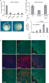
sFRPs and Fz7 act redundantly to activate Ror2. A and B, relative (rel) expression of papc analyzed by qPCR in gastrula stage 10.5 whole embryos injected dorsal-equatorially with the indicated Mo (15 ng) and synthetic mRNA (200 pg). Co, control. C, papc expression of Fz7 morphants analyzed by whole mount in situ hybridization of gastrula stage embryos. Arrowheads indicate the site of injection. D, relative expression of papc analyzed by qPCR in AC explants injected with the indicated synthetic mRNAs (500 pg of ror2/fz7 and 150 pg of wnt5a). A, B, and D show a representative with technical triplicates. This was confirmed by at least two independent experiments in different batches of X. laevis with similar results. E, confocal microscopy analysis of DMZ explants of Xenopus embryos injected dorsal-equatorially at the four-cell stage with 500 pg of Ror2-myc or 500 pg of sFRP2-HA. Endogenous Fz7 is shown in green, and overexpressed Ror2 and sFRP2 are shown in the indicated colors.
FIGURE 5.
FIGURE 5.
sFRP2 and Ror2 inhibit Fz7 mediated non-canonical Wnt signaling. A, co-IP in HEK293T cells transfected with Fz7-myc and sFRP2-HA (1 μg of each). Protein lysates were precipitated with a mouse antibody against myc or HA, respectively, and IgG as a negative control. WB analyzed with anti-HA antibody shows that sFRP2 is pulled down together with Fz7. B—D, qPCR analysis of AC explants of embryos injected with the indicated mRNAs (500 pg of n7c5/sfrp2/ror2/ror2 kd and 100 pg of wnt5a. rel, relative. E and F, ATF luciferase reporter assay of stage 12 gastrula embryos injected with the indicated synthetic mRNAs (300 pg of fz7/sfrp2/ror2/ror2 kd and 100 pg of wnt5a per embryo) and the ATF2 firefly and pRL-TK Renilla luciferase reporter constructs. B and C show the mean ± S.D. of three independent experiments. *, p<0.05; **, p<0.001 compared with controls, Student's t test. D shows a representative with technical triplicates. This was confirmed by at least two independent experiments in different batches of X. laevis with similar results. E and F show the mean of 3 ± S.E. of biological triplicates of pools of five embryos each (*, p < 0.05 to controls). This was confirmed in at least two independent experiments in different batches of X. laevis.
FIGURE 6.
FIGURE 6.
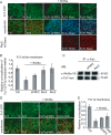
sFRP2 and Ror2 inhibit Fz7 receptor endocytosis. A and D, confocal microscopy analysis of live zebrafish embryos expressing 1 ng of mRNA of indicated constructs at 30–50% epiboly stage together with 1 ng of mRNA of the membrane marker mem-mCherry (red) or GFP-GPI (blue). Fz7-CFP (green) is present at the membrane and in endocytic vesicles. Co-expression of Wnt5a leads to enhanced internalization. sFRP2 as well as Ror2-mCherry are able to decrease Wnt5a-mediated endocytosis of Fz7-CFP. B and E, relative quantification of Fz7-CFP/membrane co-localization. Ctrl represents Fz7 alone. C, the Wnt5a-V5 binding assay was performed in HEK293T cells transfected with Fz7-myc alone or co-transfected with sFRP2-HA or Ror2-HA (1 μg of each). Each cell sample was treated with equal amounts of Wnt5a-V5 conditioned medium for 15 min before cells were lysed for rabbit anti-myc pulldown to precipitate Fz7. The samples were then analyzed on two different WBs. The WB on top was analyzed with anti-V5 antibody for the Wnt5a-V5 fraction bound to precipitated Fz7-myc. The bottom WB was analyzed with a mouse anti-myc and shows that equal amounts of Fz7-myc were precipitated in the different samples. D, co-expression of Wnt5a leads to enhanced internalization, which is repressed by full-length sFRP2 (also compare with A). The CRD of sFRP2 alone is unable to prevent Wnt5a-induced stabilization of Fz7 at the membrane but appears to increase the size of Fz7 vesicles.
FIGURE 7.
FIGURE 7.
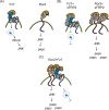
Model for selective Fz7- or Ror2-mediated activation of non-canonical Wnt signaling. A, when expressed alone, Fz7 or Ror2 receptors activate distinct non-canonical Wnt pathways stimulated by Wnt5a. B, when sFRP2 is present, Fz7 receptor endocytosis is prevented, and Fz7 signaling is inhibited, whereas Ror2 signaling is enhanced indicated by stabilized Wnt5a-Ror2 membrane complexes. C, when both receptors are present in the same cell, the Ror2-CRD acts in a similar way as sFRP2, leading to increased Ror2 activation at the expense of Fz7 signaling.

References

    1. Janda C. Y., Waghray D., Levin A. M., Thomas C., and Garcia K. C. (2012) Structural basis of Wnt recognition by Frizzled. Science 337, 59–64 - PMC - PubMed
    1. Kumar S., Žigman M., Patel T. R., Trageser B., Gross J. C., Rahm K., Boutros M., Gradl D., Steinbeisser H., Holstein T., Stetefeld J., and Özbek S. (2014) Molecular dissection of Wnt3a-Frizzled8 interaction reveals essential and modulatory determinants of Wnt signaling activity. BMC Biol. 12, 44. - PMC - PubMed
    1. Dann C. E., Hsieh J. C., Rattner A., Sharma D., Nathans J., and Leahy D. J. (2001) Insights into Wnt binding and signalling from the structures of two Frizzled cysteine-rich domains. Nature 412, 86–90 - PubMed
    1. Niehrs C. (2012) The complex world of WNT receptor signalling. Nat. Rev. Mol. Cell Biol. 13, 767–779 - PubMed
    1. Malinauskas T., and Jones E. Y. (2014) Extracellular modulators of Wnt signalling. Curr. Opin. Struct. Biol. 29, 77–84 - PubMed

Publication types

MeSH terms

LinkOut - more resources