Review
. 2016 May;39(5):300-310.
doi: 10.1016/j.tins.2016.02.006. Epub 2016 Apr 26.

NMDA Receptor Internalization by Autoantibodies: A Reversible Mechanism Underlying Psychosis?

Affiliations
Review

NMDA Receptor Internalization by Autoantibodies: A Reversible Mechanism Underlying Psychosis?

Joseph C Masdeu et al. Trends Neurosci. 2016 May.

Abstract

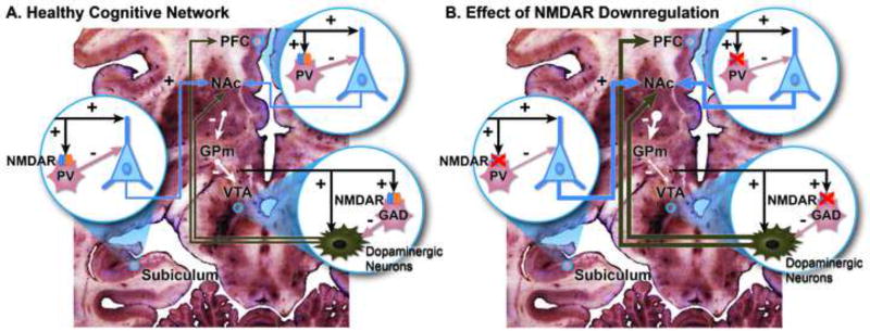
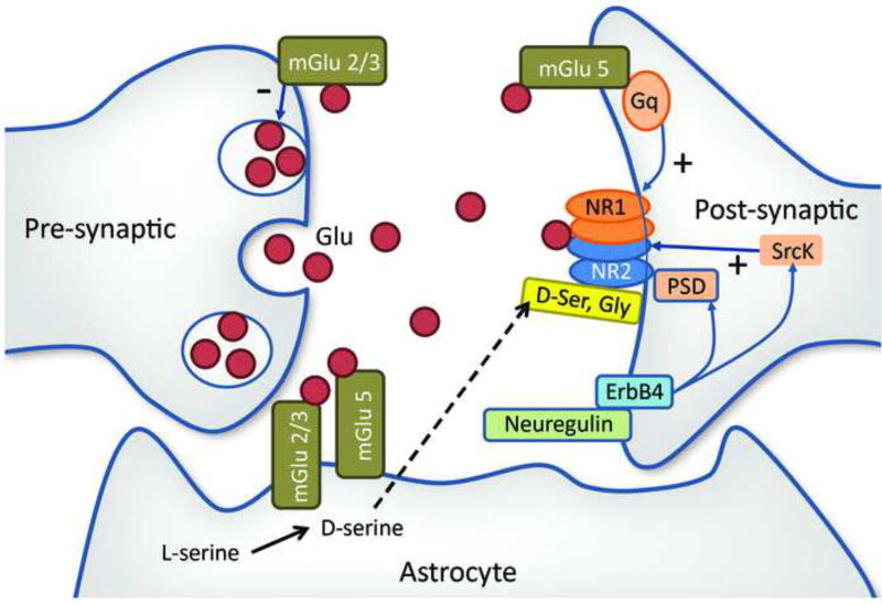
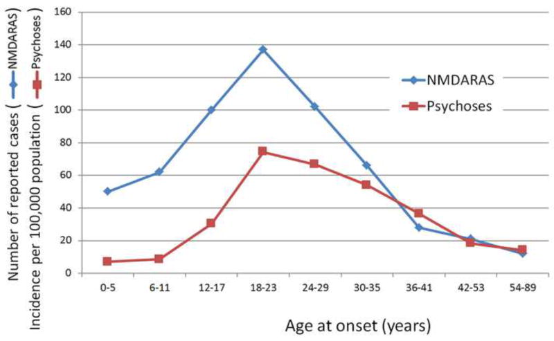
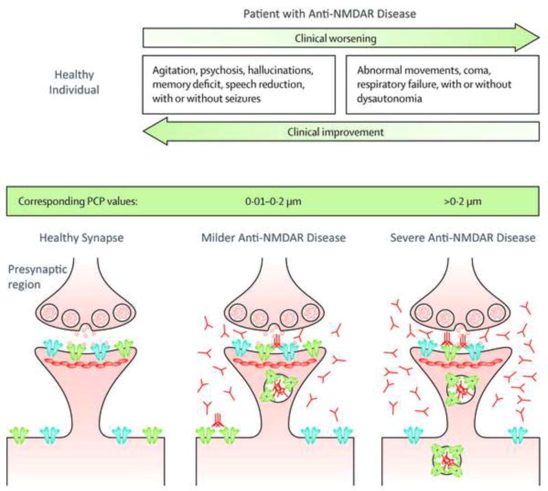
Since the early 1990s it has been postulated that hypofunction of N-methyl-d-aspartate (NMDA) receptors in brain networks supporting perception and cognition underlies schizophrenic psychosis. Recently, NMDA receptor hypofunction was described in patients with psychotic manifestations who exhibited autoantibodies binding the GluN1 subunit of the receptor, and who improved when the level of these antibodies was lowered by immunomodulation. In this disorder, NMDA receptor antibodies decrease the availability of NMDA receptors by internalizing them. In this opinion article, we review this mechanism as well as data supporting or refuting the possibility that this disorder or similar autoimmune disorders affecting synaptic proteins, which are therefore treatable with immunomodulation, could account for some cases of idiopathic psychosis. We also suggest methodological approaches to clarify this issue.

Keywords: NMDA receptor; antibodies; immunotherapy; psychosis; schizophrenia; synapsis.


Conflict of interest statement

Conflict of Interest: JCM and KFB report no conflicts of interest.

Figures

Figure 1
Figure 1
Effect of NMDA receptor (NMDAR) hypofunction. This is a simplified diagram of some of the cognitive networks that are affected by NMDAR hypofunction. Three critical nodes are shown: (1) The subiculum, effector region of the hippocampal formation; (2) the prefrontal cortex (PFC), supporting working memory and executive function; and (3) the ventral tegmental area (VTA), which houses dopaminergic neurons involved in facilitating episodic and working memory, as well as motivation. (A) At each of these nodes, glutamatergic excitatory inputs to pyramidal or dopaminergic neurons (these from multiple brain regions, prominently lateral hypothalamus [101]) provide collaterals to NMDARs on gabaergic neurons, which in their turn inhibit excessive pyramidal firing. Gabaergic interneurons in the subiculum and PFC are parvalbumin positive (PV) and, in the PFC are identified with the fast-spiking cells, critical for the generation of synchronous gamma oscillations [53, 102]. In the VTA, gabaergic interneurons also harbor NMDARs [103] and stain with glutamic acid decarboxylase (GAD); by contrast VTA PV neurons are mostly projection neurons [104]. Dopaminergic VTA neurons project to the nucleus accumbens (NAc), PFC and hippocampus (not shown). The NAc inhibits the globus pallidus medialis (GPm), which in turn inhibits tonically VTA dopaminergic neurons [10, 54]. (B) NMDAR hypofunction is associated with increased pyramidal firing, which in turn increases the inhibitory activity of the NAc over the GPm and lessens its inhibitory tone over the VTA dopaminergic neurons. As a result, there is an increased production of dopamine, as found in psychoses, which are also attended by impaired working memory associated with abnormal functioning of PV interneurons [39, 54]. Increased pyramidal firing has also been documented with NMDAR ablation restricted to the NMDARs on frontal pyramidal neurons [105].
Figure 2
Figure 2
Simplified diagram of some of the synaptic receptors known to modulate NMDARs. The excitatory neurotransmitter glutamate (Glu) is secreted at the presynaptic terminal and activates the NMDA receptor on the postsynaptic membrane. The NMDAR is a tetramer composed of two GluN1 and two NR2 subunits. It is modulated by a number of synaptic proteins. On the presynaptic membrane, metabotropic glutamate type 2/3 receptors (Glu 2/3), which bind glutamate, downregulate Glu production, while in the postsynaptic membrane metabotropic glutamate type 5 receptors (Glu 5) upregulate the NMDAR via the Gq/11 protein and phospholipase C enzyme [106]. Src kinase (Src kinase) contributes to stabilize NR2 though its action on the postsynaptic density (PSD) complex [107, 108]. Enhanced ErbB4 signaling through PSD-95 and neuregulin 1 may cause NMDAR hypofunction [109]. NMDAR activation requires the presence of glycine (Gly) or D-serine (D-ser) occupying a binding site in the GluN1 subunit [110].
Figure 3
Figure 3
Age at onset of NMDA receptor antibody synaptopathy (NMDARAS) and of schizophrenic psychosis. Graphic built with data from [20] for NMDARAS and from [111, 112] for schizophrenic psychoses.
Figure 4
Figure 4
Diagram illustrating how more severe clinical findings in patients with anti-NMDAR disease are associated with lesser NMDA receptor availability on the postsynaptic region and higher levels of brain antibodies. The clinical picture resembles that caused by phencyclidine (PCP, green bar), in which the profile of symptoms correlates with the circulating concentration of the drug. Post-synaptic receptors depicted in blue represent α-amino-3-hydroxy-5-methyl-4-isoxazolepropionic acid receptors (AMPAR), those in green N-methyl-D-aspartate receptors (NMDAR). The small round vesicles that are released in the synapse represent glutamate. Modified from Dalmau et al [74].

References

    1. Hansen KB, et al. Distinct functional and pharmacological properties of Triheteromeric GluN1/GluN2A/GluN2B NMDA receptors. Neuron. 2014;81:1084–1096. - PMC - PubMed
    1. Karakas E, Furukawa H. Crystal structure of a heterotetrameric NMDA receptor ion channel. Science. 2014;344:992–997. - PMC - PubMed
    1. Lee CH, et al. NMDA receptor structures reveal subunit arrangement and pore architecture. Nature. 2014;511:191–197. - PMC - PubMed
    1. Paoletti P, et al. NMDA receptor subunit diversity: impact on receptor properties, synaptic plasticity and disease. Nat Rev Neurosci. 2013;14:383–400. - PubMed
    1. Wyllie DJ, et al. Influence of GluN2 subunit identity on NMDA receptor function. Neuropharmacology. 2013;74:4–17. - PMC - PubMed