

Consensus Recommendations for Radiation Therapy Contouring and Treatment of Vulvar Carcinoma
- PMID: 27130794
- PMCID: PMC5189987
- DOI: 10.1016/j.ijrobp.2016.02.043
Consensus Recommendations for Radiation Therapy Contouring and Treatment of Vulvar Carcinoma
Abstract
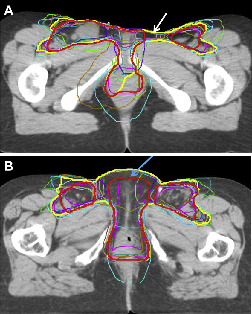
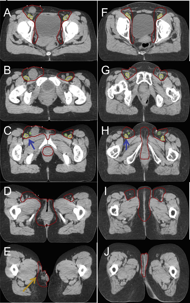
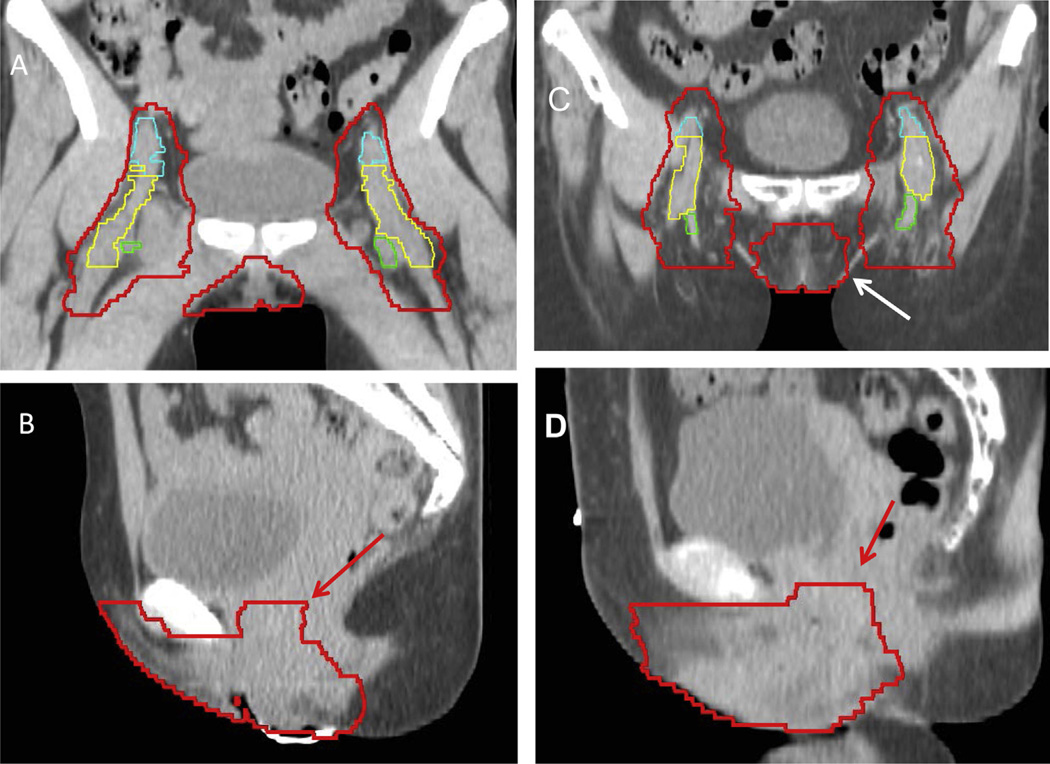
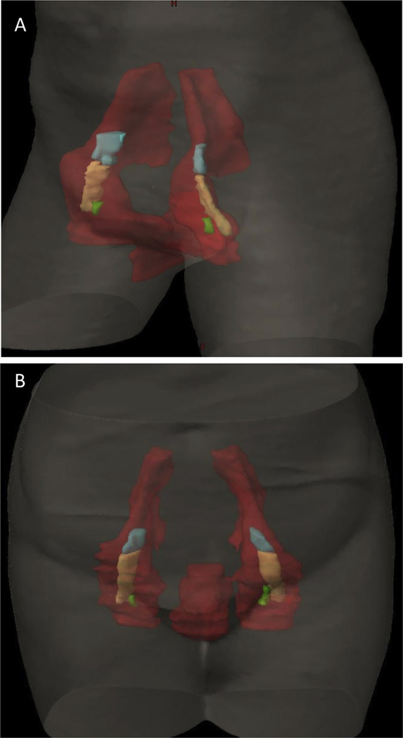
Purpose: The purpose of this study was to develop a radiation therapy (RT) contouring atlas and recommendations for women with postoperative and locally advanced vulvar carcinoma.
Methods and materials: An international committee of 35 expert gynecologic radiation oncologists completed a survey of the treatment of vulvar carcinoma. An initial set of recommendations for contouring was discussed and generated by consensus. Two cases, 1 locally advanced and 1 postoperative, were contoured by 14 physicians. Contours were compared and analyzed using an expectation-maximization algorithm for simultaneous truth and performance level estimation (STAPLE), and a 95% confidence interval contour was developed. The level of agreement among contours was assessed using a kappa statistic. STAPLE contours underwent full committee editing to generate the final atlas consensus contours.
Results: Analysis of the 14 contours showed substantial agreement, with kappa statistics of 0.69 and 0.64 for cases 1 and 2, respectively. There was high specificity for both cases (≥99%) and only moderate sensitivity of 71.3% and 64.9% for cases 1 and 2, respectively. Expert review and discussion generated consensus recommendations for contouring target volumes and treatment for postoperative and locally advanced vulvar cancer.
Conclusions: These consensus recommendations for contouring and treatment of vulvar cancer identified areas of complexity and controversy. Given the lack of clinical research evidence in vulvar cancer radiation therapy, the committee advocates a conservative and consistent approach using standardized recommendations.
Copyright © 2016 Elsevier Inc. All rights reserved.
Conflict of interest statement
All other authors report no conflicts of interest.
Figures
References
-
- National Cancer Institute Surveillance, Epidemiology, and End Results Program. SEER stat fact sheets: Vulvar cancer. [Accessed February 27, 2016]; Available at: http://seer.cancer.gov/statfacts/html/vulva.html.
-
- Hampl M, Deckers-Figiel S, Hampl JA, et al. New aspects of vulvar cancer: Changes in localization and age of onset. Gynecol Oncol. 2008;109:340–345. - PubMed
-
- Klemba A, Kukwa W, Semczuk A, et al. Vulvar cancer as a target for molecular medicine. Front Biosci. 2011;3:136–144. - PubMed
-
- Kunos C, Simpkins F, Gibbons H, et al. Radiation therapy compared with pelvic node resection for node-positive vulvar cancer: A randomized controlled trial. Obstet Gynecol. 2009;114:537–546. - PubMed
-
- Moore DH, Thomas GM, Montana GS, et al. Preoperative chemoradiation for advanced vulvar cancer: A phase II study of the Gynecologic Oncology Group. Int J Radiat Oncol Biol Phys. 1998;42:79–85. - PubMed
MeSH terms
Grants and funding
LinkOut - more resources
Full Text Sources
Other Literature Sources
Medical
