. 2016 Jun 1;25(11):2220-2233.
doi: 10.1093/hmg/ddw090. Epub 2016 Apr 30.

ERK1/2 directly acts on CTGF/CCN2 expression to mediate myocardial fibrosis in cardiomyopathy caused by mutations in the lamin A/C gene

Affiliations

ERK1/2 directly acts on CTGF/CCN2 expression to mediate myocardial fibrosis in cardiomyopathy caused by mutations in the lamin A/C gene

Maria Chatzifrangkeskou et al. Hum Mol Genet. .

Abstract

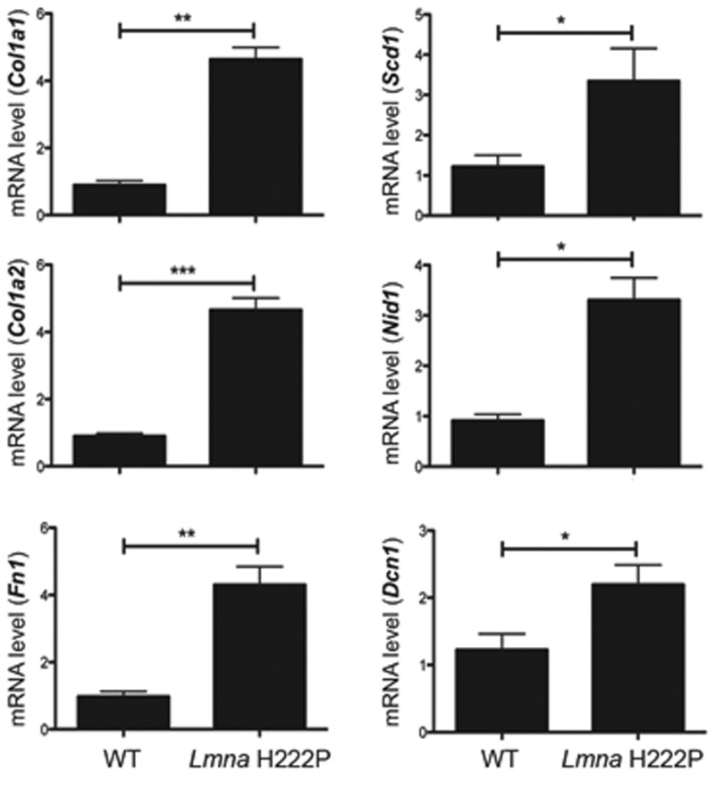
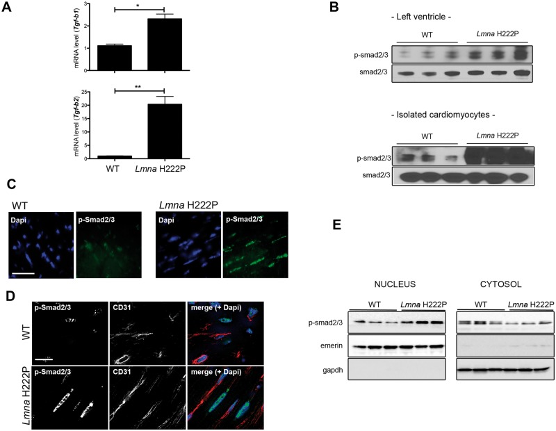
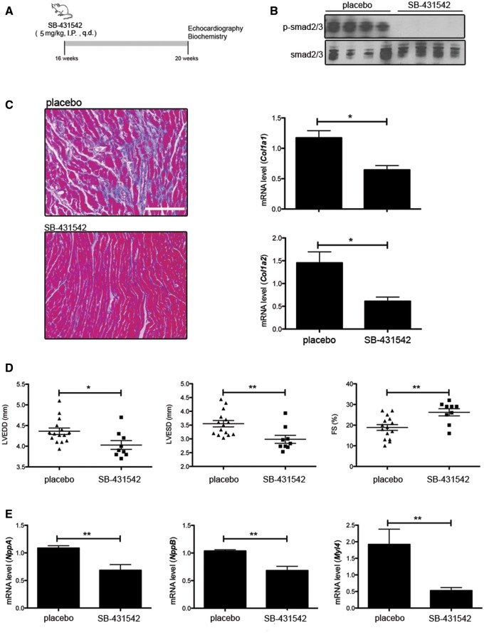
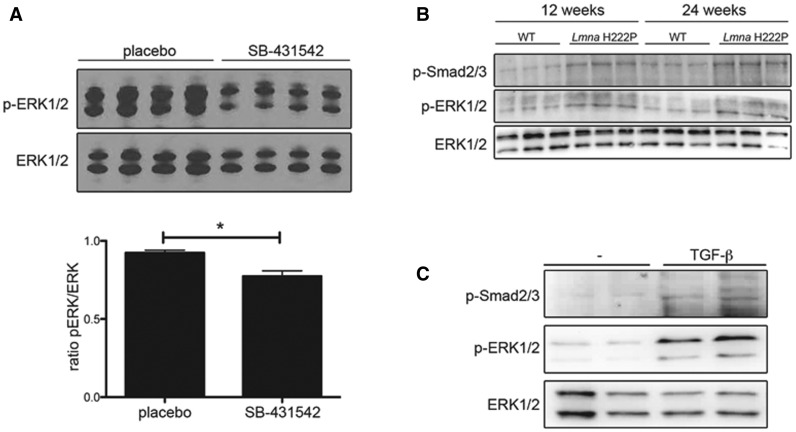
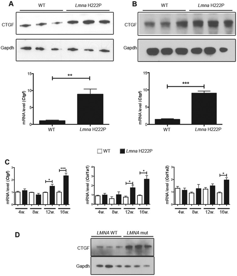
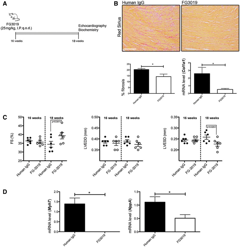
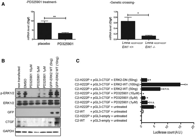
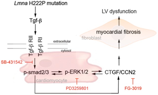
Cardiomyopathy caused by lamin A/C gene mutations (LMNA cardiomyopathy) is characterized by increased myocardial fibrosis, which impairs left ventricular relaxation and predisposes to heart failure, and cardiac conduction abnormalities. While we previously discovered abnormally elevated extracellular signal-regulated kinase 1/2 (ERK1/2) activities in heart in LMNA cardiomyopathy, its role on the development of myocardial fibrosis remains unclear. We now showed that transforming growth factor (TGF)-β/Smad signaling participates in the activation of ERK1/2 signaling in LMNA cardiomyopathy. ERK1/2 acts on connective tissue growth factor (CTGF/CCN2) expression to mediate the myocardial fibrosis and left ventricular dysfunction. Studies in vivo demonstrate that inhibiting CTGF/CCN2 using a specific antibody decreases myocardial fibrosis and improves the left ventricular dysfunction. Together, these findings show that cardiac ERK1/2 activity is modulated in part by TGF-β/Smad signaling, leading to altered activation of CTGF/CCN2 to mediate fibrosis and alter cardiac function. This identifies a novel mechanism in the development of LMNA cardiomyopathy.


Figures

Figure 1.
Figure 1.
Quantification of myocardial fibrosis in LmnaH222P/H222P mice. Cardiac expression of genes encoding proteins of the ECM in LmnaH222P/H222P mice. Bar diagrams represent relative mean mRNA (as well as standard errors of means) expression of Col1a1, Col1a2, Sdc1, Nid1, Fn1 and Dcn1 in hearts from WT mice (n = 4) and LmnaH222P/H222P mice (Lmna H222P) (n = 8). *P < 0.05, **P < 0.01, ***P < 0.001.
Figure 2.
Figure 2.
Activity of Tgf-β signaling in LmnaH222P/H222P mice. (A) Expression of genes encoding proteins of the Tgf-β signaling in heart from LmnaH222P/H222P mice. Bar graphs indicate the expression of Tgf-b1 and Tgf-b2 from WT mice (n = 3) and LmnaH222P/H222P mice (Lmna H222P) (n = 8). *P < 0.05, **P < 0.01. (B) Immunoblots showing phosphorylated Smad2/3 (p-smad2/3) and total smad2/3 in protein extracts of hearts (upper panel) and isolated cardiomyocytes (lower panel) from WT and LmnaH222P/H222P mice (Lmna H222P). Each lane contains protein extracts from a different mouse. (C) Micrographs showing p-smad2/3 labeling (upper part, Scale bar: 25 μm) of cross sections of hearts from WT compared with LmnaH222P/H222P mice (Lmna H222P). Nuclei counter-stained with 4′,6-diamidino-2-phenylindole (dapi) are also shown. (D) Micrographs showing p-smad2/3 and CD31 staining (lower part, Scale bar: 25 μm) of cross sections of hearts from WT compared with LmnaH222P/H222P mice (Lmna H222P). Nuclei counter-stained with 4′,6-diamidino-2-phenylindole (dapi) are also shown. (E) Immunoblots showing p-smad2/3 and total smad2/3 in nuclear and cytosol extracts from hearts from WT and LmnaH222P/H222P mice (Lmna H222P). Each lane contains protein extracts from a different mouse.
Figure 3.
Figure 3.
Pharmacological inhibition of Tgf-β signaling improves left-ventricular function. (A) Schematic representation of the treatment protocol of LmnaH222P/H222P mice with SB-431542. (B) Representative immunoblots using antibodies against p-smad2/3 and total smad2/3 to probe proteins extracted from hearts from LmnaH222P/H222P mice (Lmna H222P) treated with placebo or SB-431542. Each lane contains protein extracts from a different mouse. (C) Mason trichrome staining of cross sections of hearts from LmnaH222P/H222P mice (Lmna H222P) treated with placebo or SB-431542. Scale bar: 50 μm. Bar diagrams indicate the expression of Col1a1 and Col1a2 from LmnaH222P/H222P mice (Lmna H222P) treated with placebo (n = 3) or SB-431542 (n = 5). *P < 0.05. (D) Graph showing mean left ventricle end-diastolic diameter (LVEDD), mean left ventricle end-systolic diameter (LVESD) and fraction shortening (FS) in 20-week-old male LmnaH222P/H222P mice (Lmna H222P) treated with placebo (n = 15) or SB-431542 (n = 9). Values for each individual mouse receiving placebo or SB-431542 as well as standard errors of means (bars) are shown. *P < 0.05, **P < 0.01. (E) Relative expression of mRNAs from genes encoding myosin light chain (Myl4), atrial natriuretic peptide A (NppA) and brain natriuretic peptide B (NppB) in LmnaH222P/H222P mice (Lmna H222P) treated with placebo (n = 3) or SB-431542 (n = 5). Values shown are means ± standard errors. **P < 0.01.
Figure 4.
Figure 4.
Pharmacological inhibition of ERK1/2 signaling acts on Tgf-β signaling in LmnaH222P/H222P mice. Micrographs showing p-smad2/3 labeling (immunohistochemistry, Scale bar: 50 μm) of cross sections of hearts from WT compared with LmnaH222P/H222P mice (Lmna H222P) treated with placebo or PD325901. Nuclei are counter-stained with 4′,6-diamidino-2-phenylindole (dapi).
Figure 5.
Figure 5.
Tgf-β signaling acts on ERK1/2 signaling in LmnaH222P/H222P mice. (A) Immunoblots showing pERK1/2 and ERK1/2 in protein extracts of hearts from LmnaH222P/H222P mice (Lmna H222P) treated with placebo or SB-431542. Each lane contains protein extracts from a different mouse. The bar graph shows means ± standard errors values of the ratio pERK1/2 normalized to ERK1/2 from scan band densities of three immunoblots from n = 4 different mice per groups. *P < 0.05. (B) Immunoblots showing p-smad2/3, pERK1/2 and ERK1/2 in protein extracts of hearts from LmnaH222P/H222P mice (Lmna H222P) at 12 and 24 weeks of age. Each lane contains protein extracts from a different mouse. (C) Immunoblots showing p-smad2/3, pERK1/2 and ERK1/2 in protein extracts of C2C12 cells treated with or without (−) TGF-β1 for 6 h (2 nM).
Figure 6.
Figure 6.
Abnormal CTGF expression in hearts of LmnaH222P/H222P mice and human subjects with LMNA cardiomyopathy. (A) Immunoblots showing CTGF and GAPDH protein expression in hearts from WT and LmnaH222P/H222P mice. Each lane contains protein extracts from a different mouse. The bar graph represents mRNA relative expression (means ± standard errors of means) of Ctgf in hearts from WT (n = 3) and LmnaH222P/H222P (Lmna H222P) (n = 3) mice. **P < 0.01. (B) Immunoblots showing CTGF and GAPDH protein expression in isolated cardiomyocytes from WT and LmnaH222P/H222P mice. Each lane contains protein extracts from cardiomyocytes isolated from a different mouse. The bar graph represents mRNA relative expression (means ± standard errors of means) of Ctgf in cardiomyocytes from WT (n = 3) and LmnaH222P/H222P (Lmna H222P) (n = 3) mice. ***P < 0.001. (C) Validation of differential mRNA expression of Ctgf, Col1a1 and Col1a2 in hearts from WT and LmnaH222P/H222P mice. RNA was obtained from hearts of mice at 4, 8, 12, 16 weeks of age. White bars show relative RNA expression levels in hearts of WT mice and black bars in hearts of LmnaH222P/H222P mice. Values are means ± standard errors for n = 4 samples per group. *P < 0.05, ***P < 0.001. (D) Immunoblots showing CTGF and GAPDH in protein extracts of heart tissue from three human control (LMNA WT) and three individuals with dilated cardiomyopathy caused by LMNA mutations (LMNA mut).
Figure 7.
Figure 7.
Pharmacological inhibition of CTGF/CCN2 improves left-ventricular function in LmnaH222P/H222P mice. (A) Schematic representation of the treatment protocol of LmnaH222P/H222P mice with FG-3019. (B) Sirius Red staining of cross sections of hearts from LmnaH222P/H222P mice (Lmna H222P) treated with IgG or FG-3019. Scale bar: 50 μm. Left panel bar graph shows the quantification of myocardial fibrosis (means ± standard errors of means) from LmnaH222P/H222P mice (Lmna H222P) treated with immunoglobulin G (IgG) or FG-3019. *P < 0.05. Right panel shows expression of Col1a1 gene (means ± standard errors of means) in heart from LmnaH222P/H222P mice treated with IgG (n = 3) or FG-3019 (n = 3). *P < 0.05. (C) Graph showing mean LVEDD, mean LVESD and FS in 18-week old male LmnaH222P/H222P mice (Lmna H222P) treated with IgG (n = 6) or FG-3019 (n = 6). Values for each individual mouse receiving placebo or FG-3019 as well as standard errors (bars) are shown. (D) Expression of Myh7 and NppA genes in heart from LmnaH222P/H222P mice (Lmna H222P) treated with IgG (n = 3) or FG-3019 (n = 3). *P < 0.05, n.s. non-significant.
Figure 8.
Figure 8.
Effect of ERK1/2 on CTGF/CCN2 in hearts of LmnaH222P/H222P mice and C2C12 cells. (A) Expression of Ctgf gene in heart from LmnaH222P/H222P mice (Lmna H222P) treated with PD325901 (left panel) and LmnaH222P/H222P mice crossed with Erk1−/− mice (right panel). Bar diagrams indicate the expression of Ctgf (means ± standard errors of means) from LmnaH222P/H222P mice treated with placebo (n = 8) or PD325901 (n = 8) and from LmnaH222P/H222P/Erk1+/+ mice (n = 4) or LmnaH222P/H222P/Erk1−/− mice (n = 3). *P < 0.05, **P < 0.01. (B) C2C12 were treated with DMSO or PD325901 (10, 5 or 1 μM) or transfected with plasmid encoding ERK2-WT (5, 10 nM). Immunoblots showing phosphorylated ERK1/2 (p-ERK1/2), total ERK1/2, GFP and CTGF in protein extracts of C2C12 cells treated or transfected. (C) C2C12 cells stably expressing FLAG-tagged H222P-lamin A (C2-H222P) were transfected with the plasmid encoding the promoter of CTGF alone or in association with ERK2-WT (5, 10 nM) or ERK2-DN (5 nM). C2-H222P transfected with the plasmid encoding the promoter of CTGF were also treated with DMSO or PD325901 (10, 5 or 1 μM). After 48 h, luciferase activities induced by expression of CTGF were measured in cell lysates and normalized to β-gal activities obtained from a protein encoded by a co-transfected plasmid. Results are means ± standard errors of means of three experiments. *P < 0.05, n.s. non-significant (compared to C2-H222P transfected with the plasmid encoding the promoter of CTGF alone).
Figure 9.
Figure 9.
Proposed mechanism of myocardial fibrosis and cardiac dysfunction caused by Lmna mutation. Schematic representation of the sequential events of our hypothesis leading to left ventricular dysfunction in LMNA cardiomyopathy.

References

    1. Bonne G., Di Barletta M.R., Varnous S., Becane H.M., Hammouda E.H., Merlini L., Muntoni F., Greenberg C.R., Gary F., Urtizberea J.A. et al. (1999) Mutations in the gene encoding lamin A/C cause autosomal dominant Emery-Dreifuss muscular dystrophy. Nat. Genet., 21, 285–288. - PubMed
    1. Fatkin D., MacRae C., Sasaki T., Wolff M.R., Porcu M., Frenneaux M., Atherton J., Vidaillet H.J., Jr, Spudich S., De Girolami U. et al. (1999) Missense mutations in the rod domain of the lamin A/C gene as causes of dilated cardiomyopathy and conduction-system disease. N. Engl. J. Med., 341, 1715–1724. - PubMed
    1. Meune C., Van Berlo J.H., Anselme F., Bonne G., Pinto Y.M., Duboc D. (2006) Primary prevention of sudden death in patients with lamin A/C gene mutations. N. Engl. J. Med., 354, 209–210. - PubMed
    1. Ben Yaou R., Gueneau L., Demay L., Stora S., Chikhaoui K., Richard P., Bonne G. (2006) Heart involvement in lamin A/C related diseases. Arch. Mal. Coeur Vaiss, 99, 848–855. - PubMed
    1. Fontana M., Barison A., Botto N., Panchetti L., Ricci G., Milanesi M., Poletti R., Positano V., Siciliano G., Passino C. et al. (2013) CMR-verified interstitial myocardial fibrosis as a marker of subclinical cardiac involvement in LMNA mutation carriers. JACC Cardiovasc. Imaging, 6, 124–126. - PubMed