. 2016 May 19;62(4):558-71.
doi: 10.1016/j.molcel.2016.03.030. Epub 2016 Apr 28.

ATXN7L3 and ENY2 Coordinate Activity of Multiple H2B Deubiquitinases Important for Cellular Proliferation and Tumor Growth

Affiliations

ATXN7L3 and ENY2 Coordinate Activity of Multiple H2B Deubiquitinases Important for Cellular Proliferation and Tumor Growth

Boyko S Atanassov et al. Mol Cell. .

Abstract

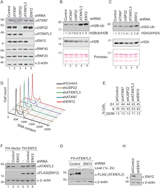
Histone H2B monoubiquitination (H2Bub1) is centrally involved in gene regulation. The deubiquitination module (DUBm) of the SAGA complex is a major regulator of global H2Bub1 levels, and components of this DUBm are linked to both neurodegenerative diseases and cancer. Unexpectedly, we find that ablation of USP22, the enzymatic center of the DUBm, leads to a reduction, rather than an increase, in global H2bub1 levels. In contrast, depletion of non-enzymatic components, ATXN7L3 or ENY2, results in increased H2Bub1. These observations led us to discover two H2Bub1 DUBs, USP27X and USP51, which function independently of SAGA and compete with USP22 for ATXN7L3 and ENY2 for activity. Like USP22, USP51 and USP27X are required for normal cell proliferation, and their depletion suppresses tumor growth. Our results reveal that ATXN7L3 and ENY2 orchestrate activities of multiple deubiquitinating enzymes and that imbalances in these activities likely potentiate human diseases including cancer.


Figures

Figure 1
Figure 1. USP22 loss does not lead to H2B deubiquitination defects in mammalian cells
A) Immunoblots to demonstrate efficient silencing of the SAGA DUBm components in 293T cells. (B and C) Depletion of ATXN7L3 and ENY2, but not USP22 or ATXN7, leads to a robust increase of bulk H2Bub1 and a moderate increase of H2Aub1 levels; compare lanes 4 and 5 to 1, 2 and 3. (D) Depletion of each SAGA DUBm component leads to a mild increase in the proportion of G1-phase cells as indicated by DNA content. (E) Cell cycle distribution percentages. (F) Depletion of ENY2 severely impacts steady state levels of ATXN7L3 (compare lanes 2 and 3). This effect can be rescued by expression of exogenous ENY2 (compare lanes 3 and 6). (G) ENY2 is required for stability of exogenous ATXN7L3. (H) Efficiency of ENY2 silencing in panel G.
Figure 2
Figure 2. ATXN7L3 interacts with USP27X and USP51
(A) Schematic of tandem FLAG-V5 affinity purification. (B) FL-V5-ATXN7L3 associated proteins identified by mass spectrometry. (C) Silver stain and immunoblot analyses to confirm interactions of ATXN7L3 with USP27X and USP51. Bands corresponding to USP27X and USP51 are indicated by black lines. (D) Immunoprecipitations of nuclear extracts (NE) using an αATXN7L3 antibody or rabbit IgG followed by immunoblots for USP27X or USP51. Input represents 5% of the NE. (E) Schematic representation of the human USP27X locus. ATG indicates the predicted translation start site of human USP27X protein (A6NNY8) and CTG indicates the experimentally discovered start site. (F) Autoradiograph of 35S-labeled, in vitro translated hUSP27X. The arrow indicates expected size of the translated protein. (G) Protein domain organization in USP22, USP27X and USP51. The sequence identity between USP22 and USP27X and between USP22 and USP51 human proteins is indicated with the brackets on the right, and the percentage identity between the ZnF-UBP domains is indicated by brackets on the left. (H) Whole cell lysates (WCL) from the indicated cell lines were immunoblotted using USP27X and USP51 specific antibodies. β-actin; loading control. (I) USP27X and USP51 display predominantly nuclear localization. The KRRK NLS sequence in USP22 is 100% conserved in USP51 and is highly similar in USP27X (top panel). USP27X-FL-V5 or USP51-FL-V5 expressing cells stained with αV5 antibody (red) and αATXN7L3 antibody (green). Yellow indicates co-localization. DAPI was used for nuclear counterstaining (blue). Scale bar = 15 μm.
Figure 3
Figure 3. USP27X and USP51 exhibit DUB activity in vivo and in vitro
(A) Schematic of the catalytic domains of USP22, USP27X, and USP51. The asterisk indicates the catalytic cysteine (C185 for USP22, C285 for USP27X and C372 for USP51), which was changed to serine (C/S) to generate catalytically dead mutants. (B) Protein levels of expressed WT or mutant USP22, USP27X, and USP51 in 293T cells. (C) Expression of the WT USPs reduces global levels of H2Bub1 in 293T cells, whereas expression of catalytically dead USPs leads to H2Bub1 increase. (D) Experimental scheme for purification of USP27X- and USP51-containing DUB modules from insect cells (top), and colloidal coomassie staining of purified complexes (bottom). (E) ATXN7L3 and ENY2 are required for USP27X and USP51 catalytic activity. USP27X or USP51 alone or together with ATXN7L3 and ENY2 were purified from Sf21 cells for use in in vitro DUB reactions using Ubiquitin-AMC as a substrate. DUB activity was measured by the resultant fluorescence intensity. To test catalytic activity towards H2Bub1, nucleosomes were purified from 293T cells stably expressing FL-V5-H2B (F) and used in in vitro DUB reactions (G).
Figure 4
Figure 4. USP22, USP27X, and UPS51 compete for ATXN7L3 and ENY2
(A) Schematic representation of the SAGA complex. USP27X and USP51 are depicted as possible substitutes of USP22. (B) USP27X and USP51 are not part of SAGA complex. FLAG-HA-GCN5 associated proteins were analyzed by immunoblot for TRRAP, ATXN7, USP22, and ATXN7L3 (confirming successful SAGA purification), and USP27X and USP51. Asterisk indicates a nonspecific band. (C) FLAG- and V5-tagged USP27X and USP51 co-immunoprecipitate ATXN7L3 and ENY2 but not GCN5 or TAF10. (D) Loss of USP22 does not lead to USP27X or USP51 incorporation into SAGA as shown by immunoblots of SAGA purified using FLAG-GCN5. (E) Increased association of USP27X with ATXN7L3 in Usp22 KO cells. Whole cell lysates from WT or Usp22 KO mES cells were immunoprecipitated using an αATXN7L3 antibody or rabbit IgG, and purified fractions were immunoblotted for USP27X. Blots were quantified using ImageJ software. (F) USP22 and USP27X compete for ATXN7L3 binding. HA-USP22, HA-USP27X, and FLAG-ATXN7L3 were mixed in in vitro binding reactions with increasing amounts of HA-USP27X (0.1, 0.5, and 1 relative to USP22 amounts, based on colloidal blue stained gel) was added as a competition for USP22. USP22-ATXN7L3 or USP27X-ATXN7L3 complexes were precipitated with αFLAG (ATXN7L3) antibody. Amounts of recovered HA-USP22 and HA-USP27X were assessed by αHA immunoblot and quantified using ImageJ software (G) Simultaneous depletion of USP22, USP27X, and USP51 leads to increased global H2Bub1 in HCT116 cells. USP22 and USP51 were depleted in WT and USP27X KO cells (bottom panels), and the H2Bub1 signal was normalized to total H2B (top panels).
Figure 5
Figure 5. USP27X and USP51 Associated Proteins
(A) USP27X and (B) USP51 containing complexes were precipitated from 293T nuclear extracts and fractionated by size on a Superose 6 column. Eluted fractions were probed for ATXN7L3 and ENY2. (C) Profile of the FLAG and V5 purified USP27X and USP51 containing complexes after elution from αV5 resin. Asterisks indicate the bait proteins. (D) List of associated proteins identified by MudPIT. Presented are proteins identified in 3 independent experiments. Average cdNASF (Normalized Distributed Spectral Abundance Factor) is shown for each protein in the purification.
Figure 6
Figure 6. USP27X and USP51 are required for proper gene expression
(A) Global levels of H2Bub1 upon depletion of USP22, USP27X, USP51, or ATXN7L3 in MCF7 cells, relative to total H2B. (B) H2Bub1 ChIP-seq analyses identified differential H2Bub1 enrichment at several loci in USP22, USP27X, or USP51 KD cells, but not in ATXN7L3 KD cells. MA plots indicate log2 fold change values (y-axis) against average log2 cpm (counts per million) values (x-axis). The green dots represent differentially enriched loci with FDR ≤ 0.05. The red lines mark log2 fold change at 1 and −1. (C) Genome browser tracks (IGV) showing H2Bub1 signal over HOXD10, HOXD12, HOXB3 and WNT5A loci upon the indicated depletions in MCF7 cells. Dashes (A, B and C) below the tracks indicate regions amplified in qPCR experiments in panel D. Scale on left (0-8) indicates the height of normalized H2Bub1 signal (see supplementary methods). (D) ChIP-qPCR of H2Bub1 levels at the indicated loci, upon depletion of USP22, USP27X, USP51, or ATXN7L3. Values (mean ± SD of three independent experiments) are expressed as a percentage of input. (E) Expression levels of WNT5A and the indicated HOX genes as measured by qRT-PCR, normalized to levels in shControl cells (set to 1). Error bars represent ± SD of three independent experiments. (F) Altered gene expression in USP22, USP27X, or USP51 depleted cells. MA plots indicate log2 fold change values (y-axis) against average log2 cpm (counts per million) values (x-axis). The red dots represent genes with FDR ≤ 0.05. The red lines mark log2 fold change at 1 and −1. (G) Venn diagram of significantly altered genes upon depletion of each USP.
Figure 7
Figure 7. USP27X and USP51 are required for xenograft tumor growth
(A) Immunoblots to demonstrate silencing efficiency of USP27X and USP51 in MCF7 cells (B) Comparison of cell growth upon silencing of USP27X or USP51. 0.3×10⁵ cells were seeded and cells were recounted after 72h. Plots show averages of 3 independent experiments. (C) Immunoblots comparing expression of WT and C285S mutant FLAG-V5-USP27X. (D) Endogenous USP27X was depleted in MCF7 cells expressing WT or C285S USP27X-V5-FL. Proliferation was measured by seeding of 0.3×10⁵ cells each and then recounting after 48h. (E) USP27X silencing efficiency in MDA-MB-231-Luc cells. (F) USP27X is required for xenograft tumor growth. 1×10⁵ MDA-MB-231-Luc cells stably expressing the indicated shRNA were injected into 8 week old NOD/SCID mice. The weights of shUSP27X expressing and control tumors are presented in (G). (H) Significant elevation of ATXN7L3 and ENY2 levels in breast cancer patients compared to healthy controls. Plot was generated based on log2 change in gene expression (708 cancer patients and 101 healthy individuals) per TCGA database. p-values in B, D, G and H are based on a Student’s t-test. (I) Model for USP22, USP27X, and USP51 function in cells. All 3 USPs require ATXN7L3 and ENY2 for activation, and in WT cells, these adapters are distributed between the different DUB complexes. Depletion of ATXN7L3 and ENY2 abolishes the activity of all 3 DUBs, leading to major increases in H2Bub1 levels. Depletion of individual USPs, however, leads to increased incorporation of ATXN7L3 and ENY2 into the remaining DUB complexes, increasing their activity.

References

    1. Atanassov BS, Dent SY. USP22 regulates cell proliferation by deubiquitinating the transcriptional regulator FBP1. EMBO Rep. 2011;12:924–930. - PMC - PubMed
    1. Atanassov BS, Evrard YA, Multani AS, Zhang Z, Tora L, Devys D, Chang S, Dent SY. Gcn5 and SAGA regulate shelterin protein turnover and telomere maintenance. Mol. Cell. 2009;35:352–364. - PMC - PubMed
    1. Bonnet J, Wang CY, Baptista T, Vincent SD, Hsiao WC, Stierle M, Kao CF, Tora L, Devys D. The SAGA coactivator complex acts on the whole transcribed genome and is required for RNA polymerase II transcription. Genes Dev. 2014;28:1999–2012. - PMC - PubMed
    1. Byrum S, Smart SK, Larson S, Tackett AJ. Analysis of stable and transient protein-protein interactions. Methods in Mol. Biol. 2012;833:143–152. - PMC - PubMed
    1. Dedio J, Jahnen-Dechent W, Bachmann M, Muller-Esterl W. The multiligand-binding protein gC1qR, putative C1q receptor, is a mitochondrial protein. J Immunol. 1998;160:3534–3542. - PubMed

MeSH terms

LinkOut - more resources