Chemical Control of Grafted Human PSC-Derived Neurons in a Mouse Model of Parkinson's Disease
- PMID: 27133795
- PMCID: PMC4892985
- DOI: 10.1016/j.stem.2016.03.014
Chemical Control of Grafted Human PSC-Derived Neurons in a Mouse Model of Parkinson's Disease
Abstract
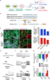
Transplantation of human pluripotent stem cell (hPSC)-derived neurons is a promising avenue for treating disorders including Parkinson's disease (PD). Precise control over engrafted cell activity is highly desired, as cells do not always integrate properly into host circuitry and can cause suboptimal graft function or undesired outcomes. Here, we show tunable rescue of motor function in a mouse model of PD, following transplantation of human midbrain dopaminergic (mDA) neurons differentiated from hPSCs engineered to express DREADDs (designer receptors exclusively activated by designer drug). Administering clozapine-N-oxide (CNO) enabled precise DREADD-dependent stimulation or inhibition of engrafted neurons, revealing D1 receptor-dependent regulation of host neuronal circuitry by engrafted cells. Transplanted cells rescued motor defects, which could be reversed or enhanced by CNO-based control of graft function, and activating engrafted cells drives behavioral changes in transplanted mice. These results highlight the ability to exogenously and noninvasively control and refine therapeutic outcomes following cell transplantation.
Copyright © 2016 Elsevier Inc. All rights reserved.
Figures


Comment in
-
Chemical Control of Grafted Human Pluripotent Stem Cell-Derived Neurons in a Mouse Parkinson Disease Model.Neurosurgery. 2016 Oct;79(4):N18-9. doi: 10.1227/01.neu.0000499710.89208.c6. Neurosurgery. 2016. PMID: 27635972 No abstract available.
Similar articles
-
Modulation of human induced neural stem cell-derived dopaminergic neurons by DREADD reveals therapeutic effects on a mouse model of Parkinson's disease.Stem Cell Res Ther. 2024 Sep 11;15(1):297. doi: 10.1186/s13287-024-03921-y. Stem Cell Res Ther. 2024. PMID: 39256801 Free PMC article.
-
Human iPSC-derived midbrain organoids functionally integrate into striatum circuits and restore motor function in a mouse model of Parkinson's disease.Theranostics. 2023 Apr 29;13(8):2673-2692. doi: 10.7150/thno.80271. eCollection 2023. Theranostics. 2023. PMID: 37215566 Free PMC article.
-
Efficiently Specified Ventral Midbrain Dopamine Neurons from Human Pluripotent Stem Cells Under Xeno-Free Conditions Restore Motor Deficits in Parkinsonian Rodents.Stem Cells Transl Med. 2017 Mar;6(3):937-948. doi: 10.5966/sctm.2016-0073. Epub 2016 Oct 14. Stem Cells Transl Med. 2017. PMID: 28297587 Free PMC article.
-
Cell therapy for Parkinson's disease: Functional role of the host immune response on survival and differentiation of dopaminergic neuroblasts.Brain Res. 2016 May 1;1638(Pt A):15-29. doi: 10.1016/j.brainres.2015.06.054. Epub 2015 Aug 1. Brain Res. 2016. PMID: 26239914 Review.
-
Embryonic and adult stem cells as a source for cell therapy in Parkinson's disease.J Mol Neurosci. 2004;24(3):353-86. doi: 10.1385/JMN:24:3:353. J Mol Neurosci. 2004. PMID: 15655260 Review.
Cited by
-
Applications of CRISPR/Cas9 for Gene Editing in Hereditary Movement Disorders.J Mov Disord. 2016 Sep;9(3):136-43. doi: 10.14802/jmd.16029. Epub 2016 Sep 21. J Mov Disord. 2016. PMID: 27667185 Free PMC article. Review.
-
Non-engineered and Engineered Adult Neurogenesis in Mammalian Brains.Front Neurosci. 2019 Feb 21;13:131. doi: 10.3389/fnins.2019.00131. eCollection 2019. Front Neurosci. 2019. PMID: 30872991 Free PMC article. Review.
-
Modulation by DREADD reveals the therapeutic effect of human iPSC-derived neuronal activity on functional recovery after spinal cord injury.Stem Cell Reports. 2022 Jan 11;17(1):127-142. doi: 10.1016/j.stemcr.2021.12.005. Stem Cell Reports. 2022. PMID: 35021049 Free PMC article.
-
Translation of WNT developmental programs into stem cell replacement strategies for the treatment of Parkinson's disease.Br J Pharmacol. 2017 Dec;174(24):4716-4724. doi: 10.1111/bph.13871. Epub 2017 Jul 9. Br J Pharmacol. 2017. PMID: 28547771 Free PMC article. Review.
-
Expansion of human midbrain floor plate progenitors from induced pluripotent stem cells increases dopaminergic neuron differentiation potential.Sci Rep. 2017 Jul 20;7(1):6036. doi: 10.1038/s41598-017-05633-1. Sci Rep. 2017. PMID: 28729666 Free PMC article.
References
-
- André VM, Cepeda C, Cummings DM, Jocoy EL, Fisher YE, William Yang X, Levine MS. Dopamine modulation of excitatory currents in the striatum is dictated by the expression of D1 or D2 receptors and modified by endocannabinoids. Eur. J. Neurosci. 2010;31:14–28. - PubMed
-
- Aravanis AM, Wang LP, Zhang F, Meltzer LA, Mogri MZ, Schneider MB, Deisseroth K. An optical neural interface: in vivo control of rodent motor cortex with integrated fiberoptic and optogenetic technology. J. Neural Eng. 2007;4:S143–S156. - PubMed
-
- Barker RA, Barrett J, Mason SL, Björklund A. Fetal dopaminergic transplantation trials and the future of neural grafting in Parkinson’s disease. Lancet Neurol. 2013;12:84–91. - PubMed
Publication types
MeSH terms
Substances
Grants and funding
LinkOut - more resources
Full Text Sources
Other Literature Sources
Medical
Research Materials

