Review
. 2016 Sep;26(9):680-693.
doi: 10.1016/j.tcb.2016.04.002. Epub 2016 Apr 28.

Structural Insights into Ring Formation of Cohesin and Related Smc Complexes

Affiliations
Review

Structural Insights into Ring Formation of Cohesin and Related Smc Complexes

Thomas Gligoris et al. Trends Cell Biol. 2016 Sep.

Abstract

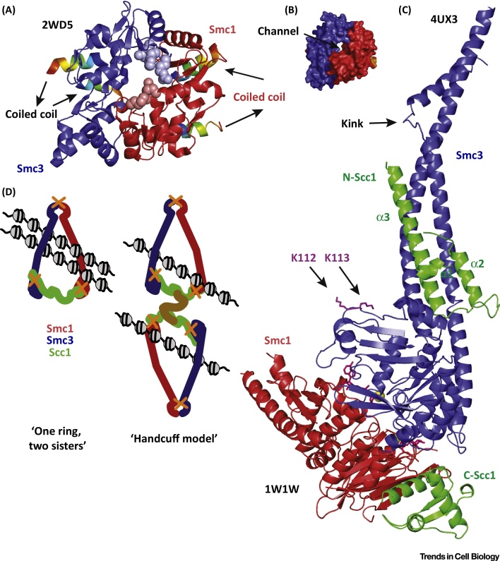
Cohesin facilitates sister chromatid cohesion through the formation of a large ring structure that encircles DNA. Its function relies on two structural maintenance of chromosomes (Smc) proteins, which are found in almost all organisms tested, from bacteria to humans. In accordance with their ubiquity, Smc complexes, such as cohesin, condensin, Smc5-6, and the dosage compensation complex, affect almost all processes of DNA homeostasis. Although their precise molecular mechanism remains enigmatic, here we provide an overview of the architecture of eukaryotic Smc complexes with a particular focus on cohesin, which has seen the most progress recently. Given the evident conservation of many structural features between Smc complexes, it is expected that architecture and topology will have a significant role when deciphering their precise molecular mechanisms.


Figures

Figure 1
Figure 1
Structure and Variations of Structural Maintenance of Chromosomes (Smc)-Kleisin Molecules. (A) Domains of a typical Smc-kleisin complex. (B) The cohesin Smc-kleisin trimer. (C) The families of Smc-kleisins. Of note are the variations within condensins. The Nse4 kleisins likely comprise a δ-kleisin group. Abbreviation: NBD, nucleotide-binding domain.
Figure 2
Figure 2
Structure of Three Cohesin Interfaces and Ring Formation. (A) Ribbon diagram of the Mus musculus structural maintenance of chromosomes 3 (Smc3; blue) and Smc1 (red) hinge interface [Protein Data Bank (PDB) 2WD5] seen from above. The entering and exiting helices of the coiled coils can be seen (rainbow colours, from blue to red following residue sequence). The side chains of the R665/K668/R669 to Alanine (Smc3) residues (light-blue spheres) and K554/K661 to Aspartate (Smc1) residues (light-blue spheres) mutated in the budding yeast charge removal experiments. (B) Surface diagram of the M. musculus hinge interface, as seen from above with the central channel. (C) Ribbon diagram of the yeast Smc3-kleisin (blue-green) interface (PDB 4UX3) combined with the yeast Smc1-kleisin (red-green) interface (PDB 1W1W). The ATP-binding pocket can be seen with ATP and Mg+2 bound (yellow sphere). The three helices of the N-terminus Scc1 and the winged helix motif of the C-terminus Scc1 interact asymmetrically with the Smcs. The acetylation patch (magenta, residues K112, K113) is distant from the Smc3-kleisin interface. (D) In vivo crosslinking data corroborate the simpler ‘one-ring, two sisters’ hypothesis rather than the ‘handcuff model’.
Figure 3
Figure 3
Cohesin Regulators and the Dynamic Life of Cohesin in the Nucleus. (A) Two main activities determine the turnover of cohesin on chromatin: Scc2-Scc4 (Nipbl-Mau2 in mammals) loads cohesin, while Pds5-Wapl-Scc3 releases cohesin from DNA. In S phase, the acetylation of structural maintenance of chromosomes 3 (Smc3) in two lysines of the nucleotide-binding domain (NBD) by Eco1 (Esco1/2) counteracts releasing, assisted by Sororin, a specific inhibitor of Wapl found in vertebrates. The cartoons are hypothetical.
Figure 4
Figure 4
Structure of the Three Components of the Cohesin-Releasing Complex. (A) Ribbon diagram of the Homo sapiens C-terminal Wapl crystal structure [Protein Data Bank (PDB) 4K6J]. Wapl (pink) comprises entirely Huntington, Elongation Factor 3, PR65/A, TOR (HEAT) repeats. A surface patch important for releasing (light red), residues mediating the interaction with the cohesin ring (red), and residues with an uncharacterised effector are highlighted. (B) Structure of the H. sapiens Scc3/SA2 (orange) interacting with the middle region of the H. sapiens Scc1-kleisin (green). A two-helix protrusion emerges from the region that Wapl and Sgo1 compete over for binding (pink). A hot spot for Scc1 binding (black surface) was determined using the D793K side chain charge reversion. (C) Ribbon diagram of the crystal structure (PDB 4XDN) of Saccharomyces cerevisiae Scc4 (magenta) interacting with the first 131 residues of Scc2 (N-Scc2, olive). Scc4 forms 13 tetratrico peptide repeats (TPR) repeats, while the middle part of N-Scc2 crosses through a channel formed by the middle domain of Scc4. A surface patch interacting with a yet unknown kinetochore component is highlighted (black). (D) Ribbon diagram of the crystal structure of the Lachancea thermotolerans Pds5 (5F0O). A small peptide of Scc1 (Lt Scc1 121–143) is shown as a green coil. A patch highlighting homologous residues corresponding to previously identified suppressors of Eco1 deletion in S. cerevisiae (most likely interacting with Wapl) are highlighted in red.
Figure 5
Figure 5
Structure of the Yeast Condensin Hinge and Coiled Coils. (A) Ribbon diagram of the crystal structure (PDB 4RSI) of the budding yeast structural maintenance of chromosomes 2 (Smc2)-Smc4 hinge with extended coiled coil domains (purple-orange). The hinge channel is almost perpendicular to the axis of the coiled coil. Residues and respective side chains used to demonstrate coiled coil contacts using thiol specific crosslinking are highlighted. (B) A combination of the known cohesin structures compiled based on known or proposed topologies. A yeast nucleosome has been added in the near background. Pds5, Scc3, and Wapl are placed at the bottom of the cohesin ring based on structural evidence. Scc2-Scc4 has been proposed to act closer to the hinge. The coiled coil domains are modelled, while all other ribbon diagrams are of published structures.

References

    1. Cobbe N., Heck M.M. The evolution of SMC proteins: phylogenetic analysis and structural implications. Mol. Biol. Evol. 2004;21:332–347. - PubMed
    1. Ames G.F., Lecar H. ATP-dependent bacterial transporters and cystic fibrosis: analogy between channels and transporters. FASEB J. 1992;6:2660–2666. - PubMed
    1. Cobbe N., Heck M.M. The evolution of ATPase activity in SMC proteins. Proteins. 2006;63:685–696. - PubMed
    1. Walker J.E. Distantly related sequences in the alpha- and beta-subunits of ATP synthase, myosin, kinases and other ATP-requiring enzymes and a common nucleotide binding fold. EMBO J. 1982;1:945–951. - PMC - PubMed
    1. Schleiffer A. Kleisins: a superfamily of bacterial and eukaryotic SMC protein partners. Mol. Cell. 2003;11:571–575. - PubMed

Publication types

MeSH terms

Substances

LinkOut - more resources