

Symmetry Breaking in an Edgeless Epithelium by Fat2-Regulated Microtubule Polarity
- PMID: 27134170
- PMCID: PMC4864504
- DOI: 10.1016/j.celrep.2016.04.014
Symmetry Breaking in an Edgeless Epithelium by Fat2-Regulated Microtubule Polarity
Abstract
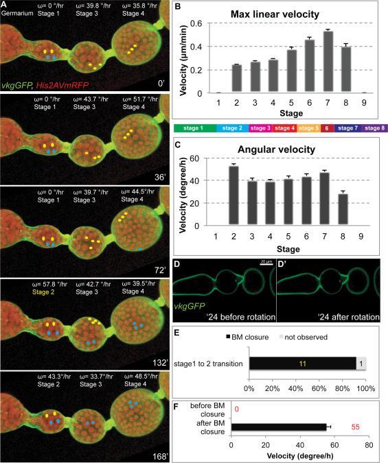
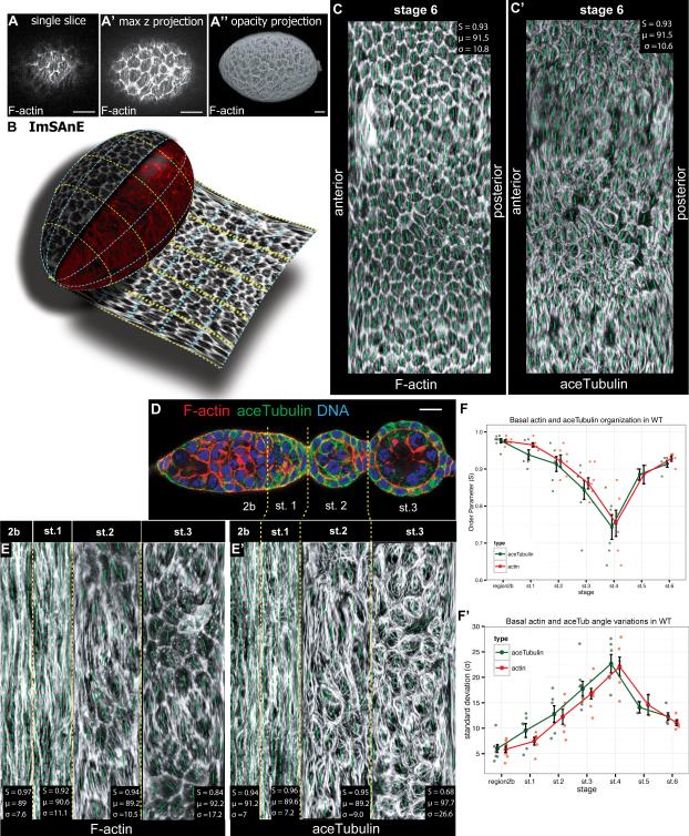
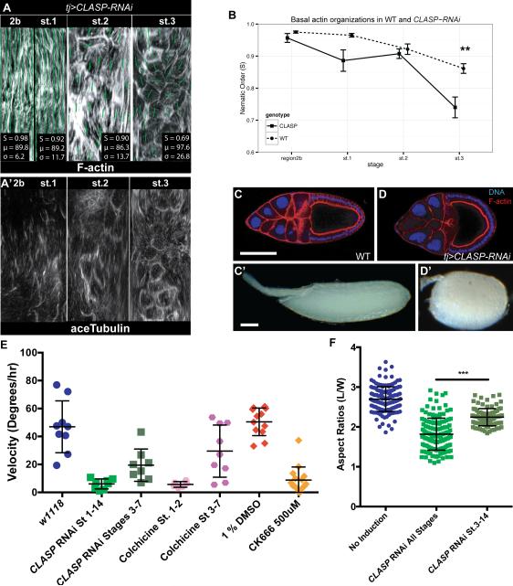
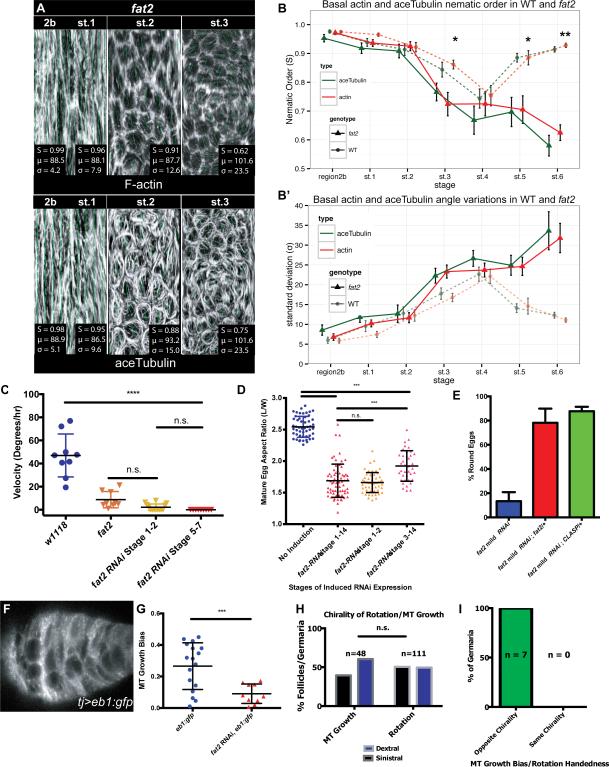
Planar cell polarity (PCP) information is a critical determinant of organ morphogenesis. While PCP in bounded epithelial sheets is increasingly well understood, how PCP is organized in tubular and acinar tissues is not. Drosophila egg chambers (follicles) are an acinus-like "edgeless epithelium" and exhibit a continuous, circumferential PCP that does not depend on pathways active in bounded epithelia; this follicle PCP directs formation of an ellipsoid rather than a spherical egg. Here, we apply an imaging algorithm to "unroll" the entire 3D tissue surface and comprehensively analyze PCP onset. This approach traces chiral symmetry breaking to plus-end polarity of microtubules in the germarium, well before follicles form and rotate. PCP germarial microtubules provide chiral information that predicts the direction of whole-tissue rotation as soon as independent follicles form. Concordant microtubule polarity, but not microtubule alignment, requires the atypical cadherin Fat2, which acts at an early stage to translate plus-end bias into coordinated actin-mediated collective cell migration. Because microtubules are not required for PCP or migration after follicle rotation initiates, while dynamic actin and extracellular matrix are, polarized microtubules lie at the beginning of a handoff mechanism that passes early chiral PCP of the cytoskeleton to a supracellular planar polarized extracellular matrix and elongates the organ.
Copyright © 2016 The Authors. Published by Elsevier Inc. All rights reserved.
Figures
Similar articles
-
Microtubule polarity predicts direction of egg chamber rotation in Drosophila.Curr Biol. 2013 Aug 5;23(15):1472-7. doi: 10.1016/j.cub.2013.06.014. Epub 2013 Jul 3. Curr Biol. 2013. PMID: 23831293
-
Epithelial rotation is preceded by planar symmetry breaking of actomyosin and protects epithelial tissue from cell deformations.PLoS Genet. 2017 Nov 27;13(11):e1007107. doi: 10.1371/journal.pgen.1007107. eCollection 2017 Nov. PLoS Genet. 2017. PMID: 29176774 Free PMC article.
-
Fat2 acts through the WAVE regulatory complex to drive collective cell migration during tissue rotation.J Cell Biol. 2016 Feb 29;212(5):591-603. doi: 10.1083/jcb.201508081. Epub 2016 Feb 22. J Cell Biol. 2016. PMID: 26903538 Free PMC article.
-
Round and round gets you somewhere: collective cell migration and planar polarity in elongating Drosophila egg chambers.Curr Opin Genet Dev. 2015 Jun;32:10-5. doi: 10.1016/j.gde.2015.01.003. Epub 2015 Feb 9. Curr Opin Genet Dev. 2015. PMID: 25677931 Free PMC article. Review.
-
Progress and challenges in understanding planar cell polarity signaling.Semin Cell Dev Biol. 2009 Oct;20(8):964-71. doi: 10.1016/j.semcdb.2009.08.001. Epub 2009 Aug 7. Semin Cell Dev Biol. 2009. PMID: 19665570 Review.
Cited by
-
Cellular and Supracellular Planar Polarity: A Multiscale Cue to Elongate the Drosophila Egg Chamber.Front Cell Dev Biol. 2021 Mar 2;9:645235. doi: 10.3389/fcell.2021.645235. eCollection 2021. Front Cell Dev Biol. 2021. PMID: 33738289 Free PMC article. Review.
-
Fat3 acts through independent cytoskeletal effectors to coordinate asymmetric cell behaviors during polarized circuit assembly.Cell Rep. 2022 Feb 1;38(5):110307. doi: 10.1016/j.celrep.2022.110307. Cell Rep. 2022. PMID: 35108541 Free PMC article.
-
Jak-Stat pathway induces Drosophila follicle elongation by a gradient of apical contractility.Elife. 2018 Feb 8;7:e32943. doi: 10.7554/eLife.32943. Elife. 2018. PMID: 29420170 Free PMC article.
-
Wave Propagation of Junctional Remodeling in Collective Cell Movement of Epithelial Tissue: Numerical Simulation Study.Front Cell Dev Biol. 2017 Jul 19;5:66. doi: 10.3389/fcell.2017.00066. eCollection 2017. Front Cell Dev Biol. 2017. PMID: 28770197 Free PMC article.
-
Configuring a robust nervous system with Fat cadherins.Semin Cell Dev Biol. 2017 Sep;69:91-101. doi: 10.1016/j.semcdb.2017.06.001. Epub 2017 Jun 9. Semin Cell Dev Biol. 2017. PMID: 28603077 Free PMC article. Review.
References
Publication types
MeSH terms
Substances
Grants and funding
LinkOut - more resources
Full Text Sources
Other Literature Sources
Molecular Biology Databases
Miscellaneous
