

Multimodality Molecular Imaging (FDG-PET/CT, US Elastography, and DWI-MRI) as Complimentary Adjunct for Enhancing Diagnostic Confidence in Reported Intermediate Risk Category Thyroid Nodules on Bethesda Thyroid Cytopathology Reporting System
- PMID: 27134564
- PMCID: PMC4809154
- DOI: 10.4103/1450-1147.176883
Multimodality Molecular Imaging (FDG-PET/CT, US Elastography, and DWI-MRI) as Complimentary Adjunct for Enhancing Diagnostic Confidence in Reported Intermediate Risk Category Thyroid Nodules on Bethesda Thyroid Cytopathology Reporting System
Abstract
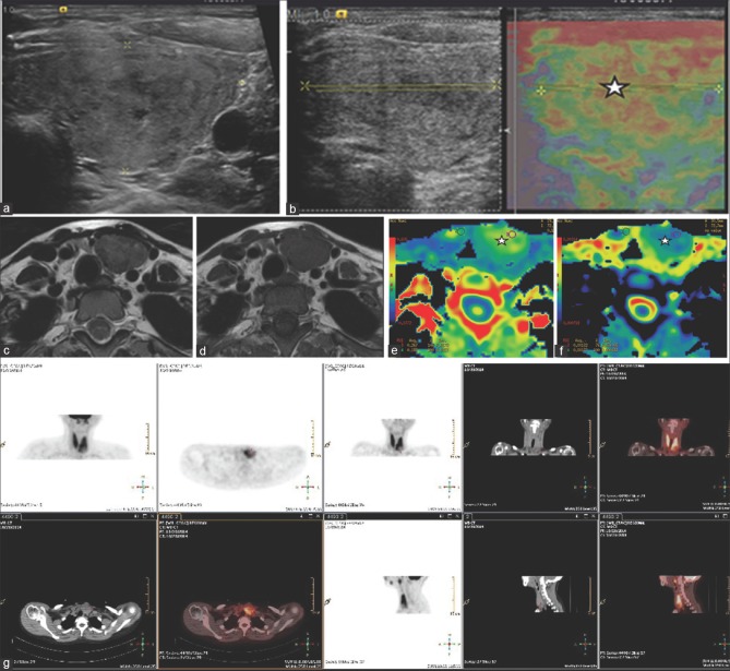
The potential complimentary role of various molecular imaging modalities [fluorodeoxyglucose-positron emission tomography/computed tomography (FDG-PET/CT), ultrasound (US)-elastography, and diffusion weighted imaging-magnetic resonance imaging (DWI-MRI)] in characterizing thyroid nodules, which have been designated as "intermediate risk category" on the Bethesda thyroid cytopathology reporting system (BTCRS), is illustrated in this communication. The clinical cases described (category III thyroid nodules on BTCRS) show the imaging features and the final diagnostic impressions rendered by the interpreting physicians with the modalities that have been independently compared in a tabular format at the end; of particular note is the high negative predictive value of these (specifically FDG-PET/CT), which could aid in enhancing the diagnostic confidence in the reported "intermediate risk category" thyroid nodules, a "gray zone" from the patient management viewpoint.
Keywords: Diffusion weighted imaging-magnetic resonance imaging (DWI-MRI); fine-needle aspiration cytology (FNAC); fluorodeoxyglucose-positron emission tomography/computed tomography (18F-FDG-PET/CT); ultrasound (US)-elastography.
Conflict of interest statement
Figures

Similar articles
-
Thyroid incidentalomas detected on 18F-fluorodeoxyglucose-positron emission tomography/computed tomography: Thyroid Imaging Reporting and Data System (TIRADS) in the diagnosis and management of patients.Surgery. 2015 Nov;158(5):1314-22. doi: 10.1016/j.surg.2015.03.017. Epub 2015 May 6. Surgery. 2015. PMID: 25958065
-
Risk stratification of thyroid nodules with Bethesda category III results on fine-needle aspiration cytology: The additional value of acoustic radiation force impulse elastography.Oncotarget. 2017 Jan 3;8(1):1580-1592. doi: 10.18632/oncotarget.13685. Oncotarget. 2017. PMID: 27906671 Free PMC article.
-
18F-Fluorodeoxyglucose Positron Emission Tomography/Magnetic Resonance in Lymphoma: Comparison With 18F-Fluorodeoxyglucose Positron Emission Tomography/Computed Tomography and With the Addition of Magnetic Resonance Diffusion-Weighted Imaging.Invest Radiol. 2016 Mar;51(3):163-9. doi: 10.1097/RLI.0000000000000218. Invest Radiol. 2016. PMID: 26784400 Free PMC article.
-
Economic Benefits and Diagnostic Quality of Diffusion-Weighted Magnetic Resonance Imaging for Primary Lung Cancer.Ann Thorac Cardiovasc Surg. 2017 Dec 20;23(6):275-280. doi: 10.5761/atcs.ra.17-00097. Epub 2017 Oct 4. Ann Thorac Cardiovasc Surg. 2017. PMID: 28978865 Free PMC article. Review.
-
Performance of 18F-FDG PET/CT in Selecting Thyroid Nodules with Indeterminate Fine-Needle Aspiration Cytology for Surgery. A Systematic Review and a Meta-Analysis.J Clin Med. 2019 Aug 28;8(9):1333. doi: 10.3390/jcm8091333. J Clin Med. 2019. PMID: 31466411 Free PMC article. Review.
Cited by
-
Diffusion magnetic resonance imaging: A molecular imaging tool caught between hope, hype and the real world of "personalized oncology".World J Radiol. 2017 Jun 28;9(6):253-268. doi: 10.4329/wjr.v9.i6.253. World J Radiol. 2017. PMID: 28717412 Free PMC article. Review.
-
The Continuing Evolution of Molecular Functional Imaging in Clinical Oncology: The Road to Precision Medicine and Radiogenomics (Part I).Mol Diagn Ther. 2019 Feb;23(1):1-26. doi: 10.1007/s40291-018-0366-4. Mol Diagn Ther. 2019. PMID: 30411216 Review.
References
-
- Cibas ES, Ali SZ. NCI Thyroid FNA State of the Science Conference. The bethesda system for reporting thyroid cytopathology. Am J Clin Pathol. 2009;132:658–65. - PubMed
-
- Hirsch D, Robenshtok E, Bachar G, Braslavsky D, Benbassat C. The implementation of the bethesda system for reporting thyroid cytopathology improves malignancy detection despite lower rate of thyroidectomy in indeterminate nodules. World J Surg. 2015 Epub ahead of print. - PubMed
-
- Basu S. Employing Bayesian approach to the intermediate risk categories of the Bethesda thyroid cytopathology reporting system: Can FDG PET/CT find a strong enough evidence-base to be practised clinically as an adjunct? Eur J Nucl Med Mol Imaging. 2014;41:2354–5. - PubMed
Publication types
LinkOut - more resources
Full Text Sources
Other Literature Sources