

The role of imaging in diagnosis and management of femoral head avascular necrosis
- PMID: 27134630
- PMCID: PMC4832402
- DOI: 10.11138/ccmbm/2015.12.3s.031
The role of imaging in diagnosis and management of femoral head avascular necrosis
Abstract
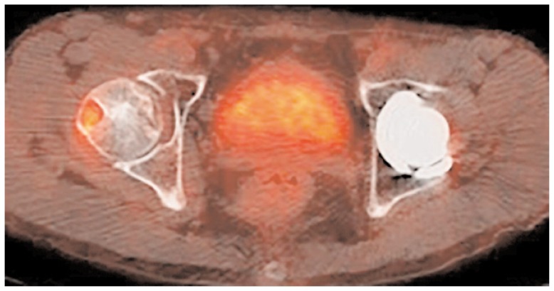
The aim of this paper is to critically review the literature documenting the imaging approach in adult Femoral Head Avascular Necrosis (FHAVN). For this purpose we described and evaluated different radiological techniques, such as X-ray, Computed Tomography (CT), Magnetic Resonance Imaging (MRI), and Nuclear Medicine. Plain films are considered the first line imaging technique due to its ability to depict femoral head morphological changes, to its low costs and high availability. CT is not a routinely performed technique, but is useful to rule out the presence of a subchondral fracture when MRI is doubtful or contraindicated. MRI is unanimously considered the gold standard technique in the early stages, being capable to detect bone marrow changes such as edema and sclerosis. It may be useful also to guide treatment and, as CT, it is a validated technique in follow-up of patients with FHAVN. Nuclear medicine imaging is mostly applied in post-operative period to detect graft viability or infective complications. More advanced techniques may be useful in particular conditions but still need to be validated; thus new research trials are desirable. In conclusion, X-ray examination is the first line approach, but lacks of sensitivity in early stage whereas MRI is indicated. CT easily depicts late stage deformation and may decrease MRI false positive results in detecting the subchondral fracture. However, the role of both Nuclear Medicine Imaging and advanced MR techniques in FHAVN still need to be investigated.
Keywords: diagnostic imaging; disease management; femur head necrosis; osteonecrosis.
Figures
Similar articles
-
F-18 fluoride positron emission tomography/computed tomography in the diagnosis of avascular necrosis of the femoral head: Comparison with magnetic resonance imaging.Indian J Nucl Med. 2016 Jan-Mar;31(1):3-8. doi: 10.4103/0972-3919.172337. Indian J Nucl Med. 2016. PMID: 26917886 Free PMC article.
-
More advantages in detecting bone and soft tissue metastases from prostate cancer using 18F-PSMA PET/CT.Hell J Nucl Med. 2019 Jan-Apr;22(1):6-9. doi: 10.1967/s002449910952. Epub 2019 Mar 7. Hell J Nucl Med. 2019. PMID: 30843003
-
Accuracy and limitations of diagnostic methods for avascular necrosis of the hip.Expert Opin Med Diagn. 2013 Mar;7(2):179-87. doi: 10.1517/17530059.2013.757592. Epub 2013 Jan 18. Expert Opin Med Diagn. 2013. PMID: 23530887 Review.
-
[CT/MRI image characteristics of iliopsoas bursitis in avascular necrosis of femoral head].Zhongguo Xiu Fu Chong Jian Wai Ke Za Zhi. 2008 Mar;22(3):295-8. Zhongguo Xiu Fu Chong Jian Wai Ke Za Zhi. 2008. PMID: 18396705 Chinese.
-
[Imaging and classification of avascular femoral head necrosis].Orthopade. 2018 Sep;47(9):729-734. doi: 10.1007/s00132-018-3615-7. Orthopade. 2018. PMID: 30083847 Review. German.
Cited by
-
Aseptic Necrosis of Femoral Head - Clinical Study.Curr Health Sci J. 2021 Apr-Jun;47(2):228-236. doi: 10.12865/CHSJ.47.02.13. Epub 2021 Jun 30. Curr Health Sci J. 2021. PMID: 34765243 Free PMC article.
-
Avascular necrosis of femoral head following COVID-19 infection.Ann Med Surg (Lond). 2023 Jul 25;85(9):4206-4210. doi: 10.1097/MS9.0000000000001098. eCollection 2023 Sep. Ann Med Surg (Lond). 2023. PMID: 37663731 Free PMC article.
-
Predicting the collapse of the femoral head due to osteonecrosis: From basic methods to application prospects.J Orthop Translat. 2017 Jan 10;11:62-72. doi: 10.1016/j.jot.2016.11.002. eCollection 2017 Oct. J Orthop Translat. 2017. PMID: 29662770 Free PMC article. Review.
-
Evaluation of bone viability in patients after girdlestone arthroplasty: comparison of bone SPECT/CT and MRI.Skeletal Radiol. 2017 Sep;46(9):1249-1258. doi: 10.1007/s00256-017-2692-8. Epub 2017 Jun 16. Skeletal Radiol. 2017. PMID: 28623409
-
Early detection of steroid-induced femoral head necrosis using 99mTc-Cys-Annexin V-based apoptosis imaging in a rabbit model.Mol Med. 2020 Dec 3;26(1):120. doi: 10.1186/s10020-020-00248-1. Mol Med. 2020. PMID: 33272196 Free PMC article.
References
-
- Lieberman JR, Berry DJ, Mont MA, Aaron RK, Callaghan JJ, Rajadhyaksha AD. Osteonecrosis of the hip: management in the 21st century. Instr Course Lect. 2003;52:337–55. - PubMed
-
- Murphey MD, Foreman KL, Klassen-Fischer MK, Fox MG, Chung EM, Kransdorf MJ. From the radiologic pathology archives imaging of osteonecrosis: radiologic-pathologic correlation. Radiographics. 2014;34( 4):1003–28. - PubMed
-
- Arlet J, Ficat RP. Forage-biopsie de la tete femorale dans I’osteonecrose primitive. Observations histopathologiques portant sur huit forances. Rev Rheum. 1964;31(31):257–64.
-
- Marcus ND, Enneking WF, Massam RA. The silent hip in idiopathic aseptic necrosis. Treatment by bone-grafting. J Bone Joint Surg Am. 1973;55(7):1351–66. - PubMed
Publication types
LinkOut - more resources
Full Text Sources
Other Literature Sources