Review
. 2016 Apr 29;5(5):51.
doi: 10.3390/jcm5050051.

Tumor Budding: The Name is EMT. Partial EMT

Affiliations
Review

Tumor Budding: The Name is EMT. Partial EMT

Alexandru Dan Grigore et al. J Clin Med. .

Abstract

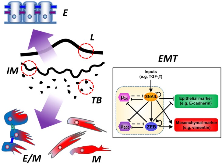
Tumor budding is a histological phenomenon encountered in various cancers, whereby individual malignant cells and/or small clusters of malignant cells are seen in the tumor stroma. Postulated to be mirror epithelial-mesenchymal transition, tumor budding has been associated with poor cancer outcomes. However, the vast heterogeneity in its exact definition, methodology of assessment, and patient stratification need to be resolved before it can be routinely used as a standardized prognostic feature. Here, we discuss the heterogeneity in defining and assessing tumor budding, its clinical significance across multiple cancer types, and its prospective implementation in clinical practice. Next, we review the emerging evidence about partial, rather than complete, epithelial-mesenchymal phenotype at the tumor bud level, and its connection with tumor proliferation, quiescence, and stemness. Finally, based on recent literature, indicating a co-expression of epithelial and mesenchymal markers in many tumor buds, we posit tumor budding to be a manifestation of this hybrid epithelial/mesenchymal phenotype displaying collective cell migration.

Keywords: EMT; cancer; epithelial-mesenchymal transition; tumor budding.


Figures

Figure 1
Figure 1
Tumor budding as a partial epithelial-mesenchymal transition. (Left) Tumor budding (TB) occurs mainly at the invasive tumoral margin (IM) which is opposite to the luminal margin (L). Studies have shown that tumor buds as a whole show a loss of epithelial markers and a gain of mesenchymal markers as compared to the main tumor mass. These features are compatible with epithelial-mesenchymal transition, which is why budding is considered to be the histological expression of the same. However, not all the cells in a tumor bud are purely mesenchymal, so budding rather mirrors a partial epithelial-mesenchymal transition. While the main tumor mass is most notably composed of purely epithelial (E) cells, the tumor buds are greatly enriched in purely mesenchymal (M) and even hybrid epithelial-mesenchymal (E/M) cells. (Inset) Epithelial-mesenchymal transition (EMT) is controlled by a core regulatory circuit composed of four players: two microRNAs, known as miR-34 (μ34, pink hexagon) and miR-200 (μ200, purple hexagon), respectively, and two transcription factors, referred to as SNAIL (orange ellipse) and ZEB1/2, which stands for Zinc finger E-box-binding homeobox-1 or -2 (ZEB, blue ellipse), respectively. The microRNAs promote the epithelial phenotype, characterized by synthesis of epithelial markers such as E-cadherin (green rounded rectangle). The transcription factors promote the mesenchymal phenotype, characterized by the synthesis of mesenchymal markers such as vimentin (red rounded rectangle). The core EMT regulatory circuit consists of two mutually inhibitory feedback loops, miR-34/SNAIL and miR-200/ZEB, respectively. Solid arrows depict transcriptional activation, with circled arrows showing self-activation. Solid bar-headed arrows depict transcriptional inhibition, with circled bar-headed arrows showing self-inhibition. Dotted bar-headed arrows represent microRNA-mediated regulation at a translational level. The relative concentrations of the four players at any given time point alter the resultant state of the core EMT regulatory circuit, thus altering the expression of epithelial and mesenchymal genes and consequently the synthesis of epithelial and mesenchymal markers. By this mechanism, the core EMT regulatory circuit governs the cellular phenotype, which can exist in (or dynamically switch between) a purely epithelial (E), a hybrid epithelial/mesenchymal (E/M), and a purely mesenchymal (M) phenotypic state. See text for more details on the relationship between EMT at a molecular scale and tumor budding at a histologic scale.

References

    1. De Smedt L., Palmans S., Sagaert X. Tumour budding in colorectal cancer: What do we know and what can we do? Virchows Arch. 2016;468:397–408. doi: 10.1007/s00428-015-1886-5. - DOI - PubMed
    1. Van Wyk H.C., Park J., Roxburgh C., Horgan P., Foulis A., McMillan D.C. The role of tumour budding in predicting survival in patients with primary operable colorectal cancer: A systematic review. Cancer Treat. Rev. 2015;41:151–159. doi: 10.1016/j.ctrv.2014.12.007. - DOI - PubMed
    1. Koelzer V.H., Langer R., Zlobec I., Lugli A. Tumor budding in colorectal cancer—Ready for diagnostic practice? Hum. Pathol. 2016;47:4–19. doi: 10.1016/j.humpath.2015.08.007. - DOI - PubMed
    1. Imai T. Growth patterns in human carcinoma. Their classification and relation to prognosis. Obstet. Gynecol. 1960;16:296–308. - PubMed
    1. Hase K., Shatney C., Johnson D., Trollope M., Vierra M. Prognostic value of tumor “budding” in patients with colorectal cancer. Dis. Colon Rectum. 1993;36:627–635. doi: 10.1007/BF02238588. - DOI - PubMed