. 2016 May 3;12(5):e1005621.
doi: 10.1371/journal.ppat.1005621. eCollection 2016 May.

Functional and Structural Mimicry of Cellular Protein Kinase A Anchoring Proteins by a Viral Oncoprotein

Affiliations

Functional and Structural Mimicry of Cellular Protein Kinase A Anchoring Proteins by a Viral Oncoprotein

Cason R King et al. PLoS Pathog. .

Abstract

The oncoproteins of the small DNA tumor viruses interact with a plethora of cellular regulators to commandeer control of the infected cell. During infection, adenovirus E1A deregulates cAMP signalling and repurposes it for activation of viral gene expression. We show that E1A structurally and functionally mimics a cellular A-kinase anchoring protein (AKAP). E1A interacts with and relocalizes protein kinase A (PKA) to the nucleus, likely to virus replication centres, via an interaction with the regulatory subunits of PKA. Binding to PKA requires the N-terminus of E1A, which bears striking similarity to the amphipathic α-helical domain present in cellular AKAPs. E1A also targets the same docking-dimerization domain of PKA normally bound by cellular AKAPs. In addition, the AKAP like motif within E1A could restore PKA interaction to a cellular AKAP in which its normal interaction motif was deleted. During infection, E1A successfully competes with endogenous cellular AKAPs for PKA interaction. E1A's role as a viral AKAP contributes to viral transcription, protein expression and progeny production. These data establish HAdV E1A as the first known viral AKAP. This represents a unique example of viral subversion of a crucial cellular regulatory pathway via structural mimicry of the PKA interaction domain of cellular AKAPs.


Conflict of interest statement

The authors have declared that no competing interests exist.

Figures
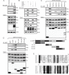
Fig 1
Fig 1. Multiple subunits of PKA are conserved targets of HAdV-5 E1A during infection.
A, B) A549 cells were infected with either WT (dl309) or ΔE1A HAdV-5 (dl312) at an MOI of 5 pfu/cell and cell lysates were harvested for co-immunoprecipitation. A) Associations between E1A and endogenous PKA subunits RIα, RIIα, and Cα are shown. B) Cells were treated with the indicated siRNAs (shown in the inset panel) prior to infection. Associations between E1A and endogenous PKA subunits RIα, RIIα, and Cα are shown. The interaction between E1A and the PKA catalytic subunit Cα required the presence of PKA regulatory subunits, indicating an indirect association. E1A’s interaction with the regulatory subunits was unaffected by the Cα knockdown. C, D) HT1080 cells were co-transfected with PKA subunits and various E1A constructs expressed as fusions to EGFP and cell lysates were harvested for co-immunoprecipitation. C) Full-length E1A proteins from 6 different HAdV species all interacted with PKA subunits RIα, RIIα, and Cα to varying degrees, with the exception of HAdV-4. D) Of the E1A fragments tested (shown in the inset panel) only the N-terminus was sufficient for interaction with PKA. This region of E1A has several regions of high amino acid sequence conservation between various HAdV species (E).
Fig 2
Fig 2. E1A contains an AKAP-like domain that is necessary and sufficient for binding PKA.
HT1080 cells were co-transfected with PKA subunits and various E1A constructs and cell lysates were harvested for co-immunoprecipitation. A) Mutational analysis using N-terminal deletion mutants of full-length E1A (shown in the inset panel) revealed that amino acids 14–28 are necessary for binding PKA subunits. B) The N-terminal region of E1A, when expressed as a fragment fused to EGFP (shown in the inset panel), was also sufficient for binding PKA subunits. This region of E1A bears amino acid similarity to a variety of known AKAPs and is predicted to contain an amphipathic α-helix (C).
Fig 3
Fig 3. E1A is predicted and confirmed to dock to PKA equivalently to a cellular AKAP.
(A,B,C) Cartoon and stick representations of the predicted E1A helix docking to Protein Kinase A regulatory subunit R1α (dimer;red). Residues represented in sticks are within 4A of each other, suggesting potential interactions. Key predicted interactions are circled. (D) Electrostatic surface representation of PKA R1α and alpha carbon tracing of E1A demonstrates limited potential electrostatic interactions, and multiple hydrophobic interactions. (E) Sequence analysis reveals the residues of PKA R1α important for AKAP binding are similar to the residues implicated in the predicted interaction with E1A. (F) Crystal structure of AKAP interacting with PKA regulatory subunit RIα (PDB ID 3IM4). AKAP structure is shown in blue, Surface electrostatics of PKA are depicted, with blue and red representing positive and negative charges respectively. (G) Sequence of the predicted E1A helix and the residues implicated in PKA interaction according to ClusPro2.0 docking. (H) E1A mutants D21K, E26K, V27K and E26A/V27A reduced interaction with RIα, whereas substitution of E25 with K, which is not predicted to alter binding, had no effect. (I) RIα mutants Q28E, L31A K32A and I35A/V36A displayed a reduced ability to bind E1A. See also S1 Fig.
Fig 4
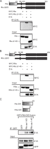
Fig 4. The D/D domains of PKA regulatory subunits are necessary and sufficient for binding E1A.
HT1080 cells were co-transfected with E1A and a variety of PKA regulatory subunit constructs and cell lysates were harvested for co-immunoprecipitation. Deletion of the D/D domain (shown in the inset panels) in either RIα (A) or RIIα (B) reduced the interaction with WT E1A. C) When expressed as EGFP fusions (shown in the inset panel) these D/D domains were sufficient for binding E1A. Images are cropped to exclude the IgG heavy chain signal.
Fig 5
Fig 5. E1A can compete with and function like an AKAP.
A) A549 cells were infected with either WT HAdV-5, ΔE1A virus, or a virus that lacks PKA-binding (Δ4–25[dl1101]) and cell lysates were harvested for co-immunoprecipitation. Interactions between the endogenous dual-specificity AKAP7 and PKA subunits was disrupted in the presence of WT E1A, but remained intact in presence of an E1A mutant unable to bind PKA. B) HT1080 cells were co-transfected with E1A, PKA, and the indicated high affinity AKAP-PKA binding inhibitors (shown in the inset panel). The binding inhibitors disrupted the E1A-PKA interactions in an isoform-specific manner. C) HT1080 cells were co-transfected with PKA and the indicated D-AKAP1 construct. An AKAP1 mutant lacking its binding for PKA was rescued by cloning in the AKAP-like sequence of HAdV-5 E1A.
Fig 6
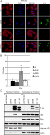
Fig 6. E1A alters PKA R1α subcellular localization during infection.
A549 cells were infected with either WT HAdV-5, ΔE1A virus or a virus mutant unable to bind PKA (Δ4–25). Cells were fixed, permeabilized and stained for confocal immunofluorescence. In the presence of WT E1A, RIα exhibited a drastic shift from exclusively cytoplasmic localization into the cell nucleus (A). This redistribution did not occur during ΔE1A or Δ4–25 infections. RIIα and Cα were not re-localized in the same manner as RIα (S2A Fig). B) Quantification of nuclear signal relative to total cellular signal. Statistically significant differences are denoted (*p<0.001) n = 50. C) Nuclear and cytoplasmic extracts from infected cells were prepared by biochemical fractionation and the presence of E1A and PKA subunits in each fraction were detected by western blot. The presence of nuclear RIα in WT-infected cells confirms the results observed by immunofluorescence. See also S2, S3 and S4 Figs.
Fig 7
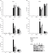
Fig 7. The interaction between E1A and PKA subunits is required for full expression of HAdV-5 E3 and E4 transcripts.
A549 cells were treated with control siRNA or siRNA specific for PKA subunits (shown in the inset panel) and infected with either WT HAdV-5 or the indicated mutants. RT-qPCR was performed with a panel of HAdV-5 early genes, normalized to GAPDH and fold change to control treated cells was plotted. Results for the HAdV-5 E1A (A), E1B (B), E2 (C), E3 (D) and E4 (E) transcription units are shown. A statistically significant decrease from control-treated cells is indicated (*p<0.05) n = 3. See also S5 and S6 Figs.
Fig 8
Fig 8. Interactions between E1A and PKA are required for full HAdV-5 progeny production.
A549 cells were treated with control siRNA or siRNA specific for PKA subunits and infected with either WT HAdV-5 or a virus encoding E1A unable to bind PKA (Δ4–25). Cells were collected at various time points up to 72 hr post-infection. Production of infectious progeny virus was quantitatively assayed by plaque formation on HEK293 cells (A). Data are shown over 36–72 hr. Growth of WT virus was decreased by knockdown of each PKA subunit. Growth of HAdV E1A Δ4–25 was not affected by knockdown of PKA regulatory subunits, but was affected by knockdown of PKA Cα (though to a lesser extent than WT HAdV-5). (B) Total viral progeny production at 60 hours post-infection when virus replication appeared to peak in most conditions. All values are represented as mean ± SEM. The statistically significant reductions in viral titres compared to control-treated cells are denoted (*p<0.01).
Fig 9
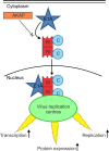
Fig 9. E1A functions as a viral AKAP.
During infection, the viral E1A protein interacts with the PKA holoenzyme. This occurs via structural mimicry of the PKA interaction domain of cellular AKAPs. As a consequence, competition occurs between the viral AKAP and cellular AKAPs for interaction with PKA. The interaction of E1A with PKA leads to a relocalization of a subset of PKA to the nucleus, likely to HAdV early gene promoters within virus replication centres. This interaction and relocalization is required for efficient viral transcription, protein expression and progeny production.

References

    1. Rozenblatt-Rosen O, Deo RC, Padi M, Adelmant G, Calderwood MA, Rolland T, et al. Interpreting cancer genomes using systematic host network perturbations by tumour virus proteins. Nature. 2012;487: 491–5. 10.1038/nature11288 - DOI - PMC - PubMed
    1. Soria C, Estermann FE, Espantman KC, O’Shea CC. Heterochromatin silencing of p53 target genes by a small viral protein. Nature. 2010;466: 1076–81. - PMC - PubMed
    1. Miyake-Stoner SJ O ’Shea CC. Metabolism goes viral. Cell Metab. 2014;19: 549–50. - PMC - PubMed
    1. Shah GA O ’Shea CC. Viral and Cellular Genomes Activate Distinct DNA Damage Responses. Cell. 2015;162: 987–1002. - PMC - PubMed
    1. Ferrari R, Pellegrini M, Horwitz GA, Xie W, Berk AJ, Kurdistani SK. Epigenetic reprogramming by adenovirus e1a. Science. 2008;321: 1086–8. 10.1126/science.1155546 - DOI - PMC - PubMed

Publication types

MeSH terms

Substances

Grants and funding