. 2016 Apr 30:16:107.
doi: 10.1186/s12870-016-0792-6.

A novel lipid transfer protein from the pea Pisum sativum: isolation, recombinant expression, solution structure, antifungal activity, lipid binding, and allergenic properties

Affiliations

A novel lipid transfer protein from the pea Pisum sativum: isolation, recombinant expression, solution structure, antifungal activity, lipid binding, and allergenic properties

Ivan V Bogdanov et al. BMC Plant Biol. .

Abstract

Background: Plant lipid transfer proteins (LTPs) assemble a family of small (7-9 kDa) ubiquitous cationic proteins with an ability to bind and transport lipids as well as participate in various physiological processes including defense against phytopathogens. They also form one of the most clinically relevant classes of plant allergens. Nothing is known to date about correlation between lipid-binding and IgE-binding properties of LTPs. The garden pea Pisum sativum is widely consumed crop and important allergenic specie of the legume family. This work is aimed at isolation of a novel LTP from pea seeds and characterization of its structural, functional, and allergenic properties.

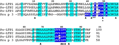
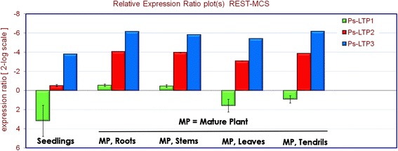
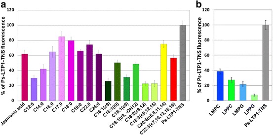
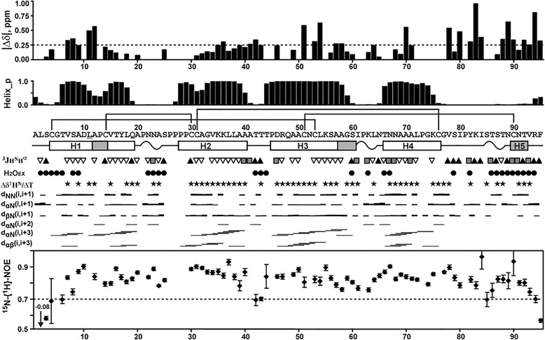
Results: Three novel lipid transfer proteins, designated as Ps-LTP1-3, were found in the garden pea Pisum sativum, their cDNA sequences were determined, and mRNA expression levels of all the three proteins were measured at different pea organs. Ps-LTP1 was isolated for the first time from the pea seeds, and its complete amino acid sequence was determined. The protein exhibits antifungal activity and is a membrane-active compound that causes a leakage from artificial liposomes. The protein binds various lipids including bioactive jasmonic acid. Spatial structure of the recombinant uniformly (13)C,(15)N-labelled Ps-LTP1 was solved by heteronuclear NMR spectroscopy. In solution the unliganded protein represents the mixture of two conformers (relative populations ~ 85:15) which are interconnected by exchange process with characteristic time ~ 100 ms. Hydrophobic residues of major conformer form a relatively large internal tunnel-like lipid-binding cavity (van der Waals volume comes up to ~1000 Å(3)). The minor conformer probably corresponds to the protein with the partially collapsed internal cavity.

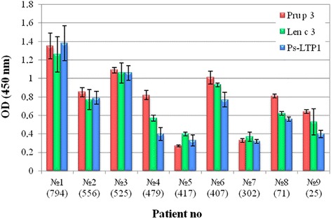
Conclusions: For the first time conformational heterogeneity in solution was shown for an unliganded plant lipid transfer protein. Heat denaturation profile and simulated gastrointestinal digestion assay showed that Ps-LTP1 displayed a high thermal and digestive proteolytic resistance proper for food allergens. The reported structural and immunological findings seem to describe Ps-LTP1 as potential cross-reactive allergen in LTP-sensitized patients, mostly Pru p 3(+) ones. Similarly to allergenic LTPs the potential IgE-binding epitope of Ps-LTP1 is located near the proposed entrance into internal cavity and could be involved in lipid-binding.

Keywords: Allergen; Antimicrobial activity; Differential gene expression; Garden pea; Heteronuclear NMR spectroscopy; Lipid binding; Lipid transfer protein; Pisum sativum; Recombinant expression; Spatial structure.


Figures

Fig. 1
Fig. 1
Amino acid sequences of novel pea LTPs and Pru p 3. The conserved residues across all sequences are highlighted in cyan. The residues participating in lipid-binding are marked with a triangles [1, 36]. The residues forming the conformational epitope of Pru p 3 are boxed in dark blue. The residues of Pru p 3 crucial for IgE-binding are asterisked. I% – percentage of sequence identity
Fig. 2
Fig. 2
Differential expression profiles of the Ps-LTP1-3 genes in various pea organs. All expression values are normalized to the reference mean of the β-tubulin gene expression. The mRNA levels in different tissue samples were calculated for each gene relatively to its expression in control pea dry seeds before germination
Fig. 3
Fig. 3
Effect of FAs and JA (a) or lysolipids (b) on the fluorescence level of the Ps-LTP1-TNS complex. FAs (18 μM) or JA (8 μM) or lysolipids (8 μM) and TNS (3.5 μM) were incubated together for 1 min and then Ps-LTP1 (4 μM) was added. Each experiment was performed in triplicate. The results are expressed as the mean values (±SD) of the percentage of the fluorescence using the Ps-LTP1-TNS complex without lipids as a control
Fig. 4
Fig. 4
NMR data define secondary structure, dynamics and conformational heterogeneity of Ps-LTP1 in solution. From top to bottom: (δΔ) Root from the sum of squared differences in 1HN and 15NH chemical shifts for the two structural forms. Differences in the 15N chemical shifts were scaled by factor 0.2. The arbitrary taken cutoff value (0.25 ppm) shows residues subjected to large-amplitude motions in ms time scale. (Helix_p) Probability of helix conformation calculated in TALOS+. The secondary structure of Ps-LTP1 is shown below the protein sequence. The α- and probable 310-helical elements are shown by white and gray bars, respectively. The helices of the protein are numbered sequentially (H1-H5). The β-turns are denoted by wavy lines. The site of Met11Leu replacement is underlined. (3JH N H α) Large (>8 Hz), small (<6 Hz) and medium (others) J-couplings are indicated by the up pointing black-filled triangles, gray-filled squares, and down pointing open triangles, respectively. (H2OEX) Amide protons which demonstrate fast exchange with water protons are shown by filled circles. The corresponding cross-peaks on the water frequency were observed in the 3D 15N-TOCSY-HSQC spectrum (τm = 80 ms). (Δδ1HN/ΔT) Black-filled stars denote amide protons with temperature gradients less than −4.5 ppb/K. NOE connectivities observed in the 80 ms 3D NOESY spectra are denoted as usual. Steady-state 15N-{1H}-NOE values are shown on the bottom of the figure. Residues displaying NOE < 0.7 are subjected to enhanced motions in ps-ns time scale
Fig. 5
Fig. 5
Spatial structure and backbone dynamics of the major structural form of Ps-LTP1 in solution. a. The sets of the best 20 structures are superimposed over the backbone atoms in regions with well-defined structure (Cys4-Cys76). The disulfide bonds are shown in orange. The helices H1-H5 are color coded. b. Ribbon representation of the Ps-LTP1 spatial structure. The ribbon is colored according to obtained dynamical NMR data (see the legend in Fig. 3). The positively charged (Arg, Lys, N-terminal amide), negatively charged (Asp, C-terminal carboxylic group), and aromatic (Phe/Tyr) side chains/moieties are in blue, red, and green, respectively. The Pro residues are shown by cyan plates. The hydrogen bonds between side chain Asn68 (magenta) and CO groups of Tyr17 and Pro21 (red cylinders), and ionic bridge (Arg47 – C-terminus) are shown by broken lines. c. Two-sided view of the Ps-LTP1 spatial structure. The side chains of hydrophobic residues (Ala/Ile/Leu/Val) which form the internal cavity are colored in yellow. The residues that form the entrance into the internal hydrophobic cavity are marked by underlined lettering. The helices H1-H5 are color coded. d, e. Two-sided views of the surfaces of Ps-LTP1 and Pru p 3 (PDB ID 2ALG, [28]) molecules, superimposed over Cα atoms of the eight conserved Cys residues. The color code is similar to one used at the other panels, except that Pro residues are colored in yellow. The two IgE epitopes (one conformational Asn35-Ala46/Ser76-Tyr79 and one sequential Ala11-Pro25) are shown on the surface of Pru p 3 by thick black lines. The Ps-LTP1 region homologues to conformational epitope of Pru p 3 is shown by dotted line. The corresponding residues on the both molecules are shown by italic and underlined lettering. Ps-LTP1 molecules shown on the panels b, right c, and left d have identical orientation. The expected entrance into internal hydrophobic cavity is shown by arrow
Fig. 6
Fig. 6
SDS-PAGE analysis of in vitro cleavage mimicking gastric and duodenal digestions of Ps-LTP1
Fig. 7
Fig. 7
Characterization of sera from patients with food allergy. Total IgE levels are specified in brackets. Data were obtained using 1:2 serum dilutions

References

    1. Carvalho ADO, Gomes VM. Role of plant lipid transfer proteins in plant cell physiology-A concise review. Peptides. 2007;28:1144–1153. doi: 10.1016/j.peptides.2007.03.004. - DOI - PubMed
    1. Park SY, Jauh GY, Mollet JC, Eckard KJ, Nothnagel EA, Walling LL, Lord EM. A lipid transfer-like protein is necessary for lily pollen tube adhesion to an in vitro stylar matrix. Plant Cell. 2000;12:151–64. - PMC - PubMed
    1. Salcedo G, Sánchez-Monge R, Barber D, Díaz-Perales A. Plant non-specific lipid transfer proteins: an interface between plant defence and human allergy. Biochim Biophys Acta. 1771;2007:781–91. - PubMed
    1. Crimi M, Astegno A, Zoccatelli G, Esposti MD. Pro-apoptotic effect of maize lipid transfer protein on mammalian mitochondria. Arch Biochem Biophys. 2006;445:65–71. doi: 10.1016/j.abb.2005.10.024. - DOI - PubMed
    1. Wang X, Zhou W, Lu Z, Ouyang Y, CS O, Yao J. A lipid transfer protein, OsLTPL36, is essential for seed development and seed quality in rice. Plant Sci. 2015;239:200–8. doi: 10.1016/j.plantsci.2015.07.016. - DOI - PubMed

Publication types

MeSH terms

LinkOut - more resources