Case Reports
. 2016 Apr 30:14:138.
doi: 10.1186/s12957-016-0887-4.

Primary intracranial neuroendocrine tumor: two case reports

Affiliations
Case Reports

Primary intracranial neuroendocrine tumor: two case reports

Hailong Liu et al. World J Surg Oncol. .

Abstract

Background: Neuroendocrine tumor originates from the diffuse neuroendocrine system. Intracranial originating is lower to 0.74 %.

Case presentation: We present two cases of primary intracranial neuroendocrine tumor A 39-year-old woman was admitted with headache, fever, polydipsia and polyuria. Biochemical and endocrinological results showed hyponatremia, hypothyroidism and hypopituitarism. MRI scans demonstrated an obviouslyenhancing lesion in seller and superseller area. Then a gross removal of tumor was achieved during the single nostril transsphenoidal approach surgery. Pathological diagnosis was high-grade small-cell neuroendocrine tumor. A 40-year-old woman presented with multiple symptoms and neurological deficit. Neuroimaging results demonstrated a huge obviously-enhancing tumor in anterior cranial fossa. Biochemical and hormone findings revealed hypokalemia, high glucose and hypercortisolemia. The intracranial surgery achieved a gross removal through a right frontal craniotomy. Pathological diagnosis was low-grade small-cell neuroendocrine tumor with immuno-negativity for ACTH.

Conclusion: The mechanism, diagnosis, and treatment of neuroendocrine tumor are still challenging.

Keywords: Anterior skull base reconstruction; Diagnosis; Ectopic ACTH syndrome; Neuroendocrine tumor; Treatment.


Figures

Fig. 1
Fig. 1
The neuroimaging results of the first patient. a CT scans demonstrated an oval high-density lesion in the sellar area and bone destruction. b T2-weighted MRI showed the lesion was slight hyperintense. c T1-weighted sagittal MRI showed the lesion was isointense. d DWI-MRI showed the lesion was iso- or hyperintensity. eg The solid part heterogeneously enhanced after contrast. h. Postoperative MRI scans (3 days later) showed a gross removal of the tumor
Fig. 2
Fig. 2
The pathological results of the first patient. a, b Specimen was comprised of different size cells with distinct atypia and proliferated actively. (HEA 100×, B 200×). ce Tumor cells were immunohistochemical positive for Syn (c), ChrA (d), and CK8 (e). f The Ki-67-labeling index was more than 90 % in the atypical cells. (IHC 100×)
Fig. 3
Fig. 3
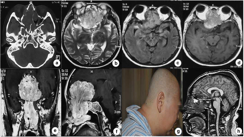
The radiological results and portraiture of the second patient. a Bone CT scans showed the neoplasm in the anterior cranial fossa eroded the bilateral sphenoid and the ethmoid sinus bone. b T2-weighted MRI showed the lesion was slight hyperintense. c The lesion showed hyperintensity in FLAIR MRI. df MRI scans demonstrated the obviously enhancing tumor infiltrated the nasal cavity, and some striped flow-empty action was found on the superior tumor border. g Preoperative picture showed the patient with fat centripetal distribution and pigmentation in the nape. h Postoperative MRI (1 week later) scans showed a gross removal of the intracranial tumor was achieved, but most part of it in the nasal cavity was removed
Fig. 4
Fig. 4
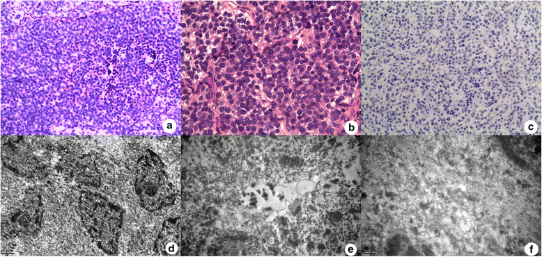
The pathological results of the second patient. a, b The tumor was comprised of small round cells with uniform nuclei and scant cytoplasm. (HEA 100×, B 400×) c The Ki-67-labeling index was less than 1 %. df Electron micrographs showed that neuroendocrine granules (NEGs) were occasionally observed. (D 10,000×, E 20,000×, F 25,000×)

References

    1. Godwin JD. Carcinoid tumors. An analysis of 2,837 cases. Cancer. 1975;36:560–9. doi: 10.1002/1097-0142(197508)36:2<560::AID-CNCR2820360235>3.0.CO;2-4. - DOI - PubMed
    1. Volante M, Rindi G, Papotti M. The grey zone between pure(neuro) endocrine and non-(neuro) endocrine tumors: a comment on concepts and classification of mixed exocrine-endocrine neoplasms. Virchows Arch. 2006;49:499–506. doi: 10.1007/s00428-006-0306-2. - DOI - PubMed
    1. Jing S, Xu Z, Wang Z, et al. Pathological category and diagnosis of neuroendocrine tumor in nonneuroendocrine system. J Clin Exp Pathol. 2009;25:548–50.
    1. Faggiano A, Mansueto G, Ferolla P, et al. Diagnosis and prognostic implication of the World Health Organization classification of neuroendocrine tumors. Endocrinol Invest. 2008;31:216–23. doi: 10.1007/BF03345593. - DOI - PubMed
    1. Waldherr C, Pless M, Maecke HR, et al. The clinical value of [90Y-DOTA]-D-Phel-Tyr3-octreotide (90Y-DOTA-TOC) in the treatment of neuroendocrine tumors: a clinical phase II study. Ann Oncol. 2001;12:941–5. doi: 10.1023/A:1011160913619. - DOI - PubMed

Publication types