. 2016 May 9;213(3):343-54.
doi: 10.1083/jcb.201603072. Epub 2016 May 2.

TANGO1 and Mia2/cTAGE5 (TALI) cooperate to export bulky pre-chylomicrons/VLDLs from the endoplasmic reticulum

Affiliations

TANGO1 and Mia2/cTAGE5 (TALI) cooperate to export bulky pre-chylomicrons/VLDLs from the endoplasmic reticulum

António J M Santos et al. J Cell Biol. .

Abstract

Procollagens, pre-chylomicrons, and pre-very low-density lipoproteins (pre-VLDLs) are too big to fit into conventional COPII-coated vesicles, so how are these bulky cargoes exported from the endoplasmic reticulum (ER)? We have shown that TANGO1 located at the ER exit site is necessary for procollagen export. We report a role for TANGO1 and TANGO1-like (TALI), a chimeric protein resulting from fusion of MIA2 and cTAGE5 gene products, in the export of pre-chylomicrons and pre-VLDLs from the ER. TANGO1 binds TALI, and both interact with apolipoprotein B (ApoB) and are necessary for the recruitment of ApoB-containing lipid particles to ER exit sites for their subsequent export. Although export of ApoB requires the function of both TANGO1 and TALI, the export of procollagen XII by the same cells requires only TANGO1. These findings reveal a general role for TANGO1 in the export of bulky cargoes from the ER and identify a specific requirement for TALI in assisting TANGO1 to export bulky lipid particles.


Figures

Figure 1.
Figure 1.
Chimeric Mia2/cTAGE5 (TALI) is structurally similar to TANGO1 and is expressed in the small intestine and liver, together with TANGO1 and ApoB. (A) Scheme depicting the domains of full-length TANGO1, cTAGE5, Mia2, and TALI. Each domain corresponds to the stretch of amino acid sequence represented by the numbers above it. (B) cDNAs from different human tissues screened for expression of TANGO1, TALI, ApoB, or the housekeeping gene GAPDH. Lu, lung; Le, leukocyte; Ov, ovary; Te, testis; S.I, small intestine; Co, colon; Sp, spleen; Th, thymus; Br, brain; He, heart; S.M, skeletal muscle; Pl, placenta; Pa, pancreas; Ki, kidney; Li, liver; Pr, prostate. The rows corresponding to the small intestine and liver profiles are highlighted; n = 3.
Figure 2.
Figure 2.
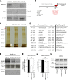
Knockout of TANGO1 or TALI impairs secretion of ApoB-containing lipid particles. (A) Western blot of differentiated parental and TANGO1- and TALI-CRISPR KO Caco-2 cells (TANGO1-KO and TALI-KO). Protein lysates were separated on a 6% SDS-PAGE, transferred onto a nitrocellulose membrane, and probed by Western blotting with anti-TANGO1 or anti-cTAGE5 antibodies; n = 3. (B) Scheme depicting the methodology used to differentiate and induce chylomicron secretion in Caco-2 cells. (C) Silver-stained gel showing proteins secreted by parental, TANGO1-KO, and TALI-KO differentiated Caco-2 cells after induction. The red arrow points to the band excised and subjected to mass spectrometry analysis; n = 3. (D) Sequences of the 30 unique peptides corresponding to ApoB as a result of mass spectrometry analysis. (E) ApoB and apoE secretion was measured by Western blotting probing supernatants collected after 5 h of induction with FCS-free medium with oleic acid. Intensities of signal in the supernatant were recorded by densitometry. Sec31 was used as a lysis control; n = 6. (F and G) Secreted ApoB and apoE levels were measured by densitometry quantification and normalized to parental cells; error bars: SEM. *, P < 0.05; **, P < 0.01; N.S, not statistically significant. (H) Total RNA was extracted from differentiated parental, TANGO1-KO, or TALI-KO Caco-2 cells, and cDNA was produced by RT-PCR and amplified to assess expression of ApoB, MTP, or the housekeeping gene GAPDH; n = 3.
Figure 3.
Figure 3.
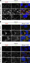
ApoB is targeted by autophagy in the absence of TANGO1 or TALI. (A and B) Differentiated parental, TANGO1-KO, or TALI-KO Caco-2 cells, induced for secretion of chylomicrons, were fixed with methanol and immunostained for ApoB (red) and LAMP1 or LC3 (green). DAPI is shown in blue. (C) Parental, TANGO1-KO, or TALI-KO HepG2 cells, induced for secretion of VLDLs, were fixed with methanol and immunostained for ApoB (red) and LC3 (green). DAPI is shown in blue. Bars, 10 µm. n = 4.
Figure 4.
Figure 4.
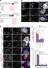
KO of TANGO or TALI blocks pre-chylomicrons and pre-VLDLs at the ER upon treatment with wortmannin. (A) Scheme depicting the methodology used to differentiate and induce chylomicron or VLDL secretion in Caco-2 or HepG2 cells, respectively, while inhibiting autophagy. (B) ApoB secretion was assessed by Western blot by probing supernatants collected after 5 h of incubation with FCS-free medium containing oleic acid in the presence or absence of the autophagy inhibitor wortmannin; intensities of signal in the supernatant were recorded by densitometry. n = 3. (C) Secreted ApoB levels with or without the autophagy inhibitor wortmannin were measured by densitometry quantification. Error bars: SEM; n = 3. (D) Differentiated parental, TANGO1-KO, or TALI-KO Caco-2 cells, induced for secretion of chylomicrons with autophagy inhibition by wortmannin, were fixed with methanol and immunostained for ApoB (red) and PDI (green). DAPI is shown in blue. Bars, 10 µm. (E) Colocalization quantifications were calculated by Manders correlation coefficient by measuring the overlap between the green and the red channels. Error bars: SEM; **, P < 0.01; ***, P < 0.001; ≥100 cells per experiment; n = 3. (F) Differentiated parental, TANGO1-KO, or TALI-KO Caco-2 cells, induced for secretion of chylomicrons with autophagy inhibition by wortmannin, were fixed with methanol and immunostained for apoE (red) and BIP (green). DAPI is shown in blue. Bars, 10 µm. (G) Parental, TANGO1-KO, or TALI-KO HepG2 cells, induced for secretion of VLDLs with autophagy inhibition by wortmannin, were fixed with methanol and immunostained for ApoB (red) and PDI (green). DAPI is shown in blue. Bars, 10 µm. (H) Colocalization quantifications were calculated by Manders correlation coefficient by measuring the overlap between the green and the red channels. Error bars: SEM; **, P < 0.01; ***, P < 0.001; ≥100 cells per experiment; n = 3.
Figure 5.
Figure 5.
KO of TANGO, but not TALI, blocks procollagen XII export from the ER. (A) Scheme depicting the methodology used to differentiate Caco-2 cells and induce secretion of collagens. (B) Lysates and supernatants of differentiated parental, TANGO1-KO, or TALI-KO Caco-2 cells, induced with ascorbic acid for 20 h, underwent Western blotting for collagen XII. Intensities of signal in the supernatant were recorded by densitometry. (C) Ratio of external versus internal collagen XII normalized to parental cells. Error bars: SEM. (D) Differentiated parental, TANGO1-KO, or TALI-KO Caco-2 cells, induced with ascorbic acid for 20 h, were fixed with methanol and immunostained for collagen XII (red) and PDI (green). DAPI is shown in blue. Bars, 10 µm. n = 3.
Figure 6.
Figure 6.
TANGO1 and TALI form a complex with ApoB at ER exit sites. (A) Caco-2 cells infected with lentivirus carrying TALI-EGFP or HepG2 transfected with TALI-EGFP were fixed with methanol and immunostained for EGFP (green) and TANGO1 (red). Bars: 10 µm; (boxes) 2.5 µm. (B) Lysates from HepG2 cells induced for VLDL secretion were incubated with no antibody (Ab; control) or with anti-TANGO1 or anti-MIA2 antibodies and then with protein A–Sepharose beads. Precipitates underwent Western blotting (WB) for TANGO1 and cTAGE5. The input sample corresponded to 10% of the total used for the immunoprecipitation. (C) Differentiated Caco-2 cells, induced for secretion of chylomicrons, were fixed with methanol and immunostained for ApoB (red) and TANGO1 or TALI/cTAGE5 (green). DAPI is shown in blue. Bars: 10 µm; (boxes) 1.25 µm. (D) Lysates from HepG2 cells induced for VLDL secretion were incubated with no antibody (control) or with anti-TANGO1, anti-cTAGE5, or anti-MIA2 antibodies and then with protein A–Sepharose beads. Precipitates underwent Western blotting for ApoB. The input sample corresponded to 10% of the total used for the immunoprecipitation. n = 3.
Figure 7.
Figure 7.
TANGO1 and TALI are both required for recruitment of ApoB to ER exit sites. (A) Scheme depicting the methodology used to differentiate and induce chylomicron secretion in Caco-2 while inhibiting ER export with BFA. (B) Differentiated Caco-2 cells, induced for secretion of chylomicrons and treated with BFA, were fixed with methanol and immunostained for ApoB (red) and Sec31, TANGO1, or ERGIC-53 (green). Boron-dipyrromethene (BODIPY) was used to visualize lipid droplets (blue). DAPI is shown in gray. Bars, 5 µm. (C) Scheme depicting the methodology used to differentiate and induce chylomicron secretion in Caco-2 while inhibiting autophagy and inhibiting ER export with BFA. (D) Differentiated parental, TANGO1-KO, or TALI-KO Caco-2 cells, induced for secretion of chylomicrons and treated with BFA or with wortmannin and BFA, were fixed with methanol and immunostained for ApoB (red) and Sec31 (green). BODIPY was used to visualize lipid droplets (blue). DAPI is shown in gray. Bars, 5 µm. (E) Colocalization quantifications were calculated by Manders correlation coefficient by measuring the overlap between the green and the red channels. Error bars: SEM; ***, P < 0.001; ≥100 cells per experiment; n = 3. (F) The number of ApoB crescents was counted by immunofluorescence microscopy, and counts were divided by the total number of cells per field of view. Error bars: SEM; ***, P < 0.001; N.S, not statistically significant; ≥100 cells per experiment; n = 3.
Figure 8.
Figure 8.
Model for the export of pre-chylomicrons/VLDLs from the ER. TANGO1 and TALI interact with ApoB directly or through an adaptor protein in the lumen of the ER to bring pre-chylomicrons/VLDLs to ER exit sites. On the cytoplasmic side, the TEER domain of TANGO1 recruits ERGIC membranes that fuse to the ER exit site that is now enriched in pre-chylomicrons/VLDLs. Binding of the proline-rich domain (PRD) of TANGO1 and TALI on the cytoplasmic side to Sec23/24 prevents Sec13/31 recruitment, allowing continuous fusion of ERGIC membranes that result in the growth of a mega-bud that is packed with pre-chylomicrons/VLDLs. Fission is triggered, and a mega carrier, big enough to accommodate bulky pre-chylomicrons/VLDLs, is released from the ER.

Comment in

References

    1. Barlowe C., and Schekman R.. 1993. SEC12 encodes a guanine-nucleotide-exchange factor essential for transport vesicle budding from the ER. Nature. 365:347–349. 10.1038/365347a0 - DOI - PubMed
    1. Bernales S., Schuck S., and Walter P.. 2007. ER-phagy: Selective autophagy of the endoplasmic reticulum. Autophagy. 3:285–287. 10.4161/auto.3930 - DOI - PubMed
    1. Blommaart E.F., Krause U., Schellens J.P., Vreeling-Sindelárová H., and Meijer A.J.. 1997. The phosphatidylinositol 3-kinase inhibitors wortmannin and LY294002 inhibit autophagy in isolated rat hepatocytes. Eur. J. Biochem. 243:240–246. 10.1111/j.1432-1033.1997.0240a.x - DOI - PubMed
    1. Bolte S., and Cordelières F.P.. 2006. A guided tour into subcellular colocalization analysis in light microscopy. J. Microsc. 224:213–232. 10.1111/j.1365-2818.2006.01706.x - DOI - PubMed
    1. Brodsky J.L., and Fisher E.A.. 2008. The many intersecting pathways underlying apolipoprotein B secretion and degradation. Trends Endocrinol. Metab. 19:254–259. 10.1016/j.tem.2008.07.002 - DOI - PMC - PubMed

MeSH terms