. 2016 May 3;12(5):e1006023.
doi: 10.1371/journal.pgen.1006023. eCollection 2016 May.

The Caenorhabditis elegans Protein FIC-1 Is an AMPylase That Covalently Modifies Heat-Shock 70 Family Proteins, Translation Elongation Factors and Histones

Affiliations

The Caenorhabditis elegans Protein FIC-1 Is an AMPylase That Covalently Modifies Heat-Shock 70 Family Proteins, Translation Elongation Factors and Histones

Matthias C Truttmann et al. PLoS Genet. .

Abstract

Protein AMPylation by Fic domain-containing proteins (Fic proteins) is an ancient and conserved post-translational modification of mostly unexplored significance. Here we characterize the Caenorhabditis elegans Fic protein FIC-1 in vitro and in vivo. FIC-1 is an AMPylase that localizes to the nuclear surface and modifies core histones H2 and H3 as well as heat shock protein 70 family members and translation elongation factors. The three-dimensional structure of FIC-1 is similar to that of its human ortholog, HYPE, with 38% sequence identity. We identify a link between FIC-1-mediated AMPylation and susceptibility to the pathogen Pseudomonas aeruginosa, establishing a connection between AMPylation and innate immunity in C. elegans.


Conflict of interest statement

The authors have declared that no competing interests exist.

Figures
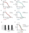
Fig 1
Fig 1. AMPylation plays a role in susceptibility to P. aeruginosa infections.
AMPylation levels have no influence on aging: wild type, fic-1(n5823) and FIC-1[E274G](nIs733) animals were kept at either 20 C (A and B) or 25 (C and D) and survival was scored every other day. Depicted n refers to number of animals at experiment initiation; number in brackets represents total counted dead events. (E) AMPylation has no consequences on pathogen avoidance: L4 nematodes were placed in the center of a P. aeruginosa loan and animal localization was scored after 24 hours. (F) FIC-1[E274G](nIs733) increases while fic-1(n5823) decreases pathogen tolerance: L4 animals were place in the center of a P. aeruginosa loan and nematode survival was scored once per day until last animal vanished. Depicted n refers to number of animals at experiment initiation; number in brackets represents total counted dead events. Representative replica shown. P-values (Gehan-Breslow-Wilcoxon test) as compared to N2 wild type control: N2. vs fic-1(n5823): 0.046; N2 vs. fic-1(n5823, nIs734) rescue: 0.009; N2 vs. FIC-1[E274G](nIs733): 0.042; fic-1(n5823) vs. fic-1(n5823, nIs734) rescue or FIC-1[E274G](nIs733): <0.0001. Additional independent replica are depicted in S7 Fig.
Fig 2
Fig 2. Hyper- or hypo-AMPylation has no apparent consequences on nematode viability and response to acute or chronic ER stress.
(A) AMPylation has no influence on development under acute ER stress: eggs were transferred to OP50 plates containing different concentrations of tunicamycin to induce acute ER stress. Embryo development was scored. Average of three independent experiments shown here. (B) AMPylation has no influence on development under chronic ER stress: eggs were transferred to P. aeruginosa plates to induce chronic ER stress. Embryo development was scored. Average of three independent experiments shown here. (C) and (D) development assay under chronic ER stress: eggs of indicated lines were transferred to P. aeruginosa plates to induce chronic ER stress. Embryo development was scored. Average of three independent experiments shown here.
Fig 3
Fig 3. FIC-1 is enriched in the adult germline as well as nematode embryos and localizes to the nuclear membrane.
(A) FIC-1 is expressed ubiquitously albeit at low levels: FIC-1 expression pattern as detected by smFISH analysis; samples stained with DAPI (left panel) and smFISH probe (middle panel). Right panel shows merged image where nuclei are represented in blue and smFISH signal in yellow. Distinct representative developmental stages shown here (B) FIC-1 is enriched in the adult germline and embryos: FIC-1 expression pattern as detected by smFISH analysis; samples stained with DAPI (left panel) and smFISH probe (middle panel). Right panel shows merged image where nuclei are represented in blue and smFISH signal in yellow. (C) FIC-1 preferentially localizes to the nuclear envelope/ER: embryos over-expressing HA-tagged FIC-1 were stained with indicated antibodies and dyes and FIC-1 localization was analyzed by confocal microscopy. (D) FIC-1 preferentially localizes to the nuclear envelope/ ER: sub-cellular fraction of C. elegans embryos. Individual fractions probed with indicated antibodies for enrichment of tested proteins. Nuc: nuclear fraction; ER: ER fraction; Cyt: cytosolic fraction.
Fig 4
Fig 4. FIC-1 is an AMPylase.
(A) FIC-1 exhibits auto-AMPylation activity: Recombinant FIC-1, FIC-1 E274G or FIC-1 H404A was incubated with α 33P-ATP for an hour and incorporation of label was assessed by SDS-PAGE and autoradiography. (B) FIC-1 accepts different nucleotide substrates: FIC-1 E274G or HYPE E234G were incubated with respective α 33P-labeled nucleotides for one hour at room temperature and sample autoradiography was assessed. (C) FIC-1 E274G AMPylates histone H3: Recombinant FIC-1, FIC-1 E274G or FIC-1 H404A was incubated with α 33P-ATP for an hour at which point histone H3 was added and the mixture was incubated for an additional hour. Incorporation of label was assessed by SDS-PAGE and autoradiography. (D) FIC-1 E274G/T476A and FIC-1 E274G/T352A are fully active: Recombinant FIC-1 E274G, FIC-1 E274G/T476A and FIC-1 E274G/T352A was incubated with α 33P-ATP for an hour at which point histone H3 was added and the mixture was incubated for an additional hour. Incorporation of label was assessed by SDS-PAGE and autoradiography. (E) FIC-1 AMPylates core histones H2 and H3 but not H4: Recombinant FIC-1, FIC-1 E274G or FIC-1 H404A was incubated with α 33P-ATP for an hour at which point purified histone substrates were added and the mixture was incubated for an additional hour. Incorporation of label was assessed by SDS-PAGE and autoradiography.
Fig 5
Fig 5. FIC-1 structure, domains and dimer interface.
(A) Ribbon representation of FIC-1 dimer, with individual domains colored in a single monomer. (B) Cartoon representation of FIC-1; the dimer interface is highlighted in the inset where key side chain and backbone contacts are shown. (C) Surface representation of FIC-1 monomer and ribbon representation of a second monomer, the ATP binding site is highlighted with a white asterisk. (D) Surface representation of FIC-1 monomer; coloring is based on conservation from an alignment between Fic-domain containing proteins. Dimerization interface is outlined in black. (E) Size exclusion chromatogram showing elution profiles of FIC-1 wildtype (wt), and FIC-1 I298D. Elution volumes of standards are highlighted with arrows. (F) Monomeric FIC-1 E274G/I298D exposes reduced AMPylation activity: FIC-1 E274G or FIC-1 E274G/I298D were pre-incubated with α 33P-ATP for an hour before histone H3 was added and the mixture was incubated for another hour. Incorporation of label was assessed by SDS-PAGE and autoradiography. (G) Quantification of histone H3 and self-AMPylation (inlet). Data shown represents the average of two independent replica. * = p-value < 0.01 (t-test).
Fig 6
Fig 6. FIC-1 AMPylates conserved heat shock 70 family proteins and translation elongation factors.
(A) Identification of new FIC-1 targets by mass spectrometry. (B) Validation of novel FIC-1 targets: Recombinant FIC-1 E274G was incubated with α 33P-ATP for an hour at which point substrates (histone H3, HSP-1 or eEF-1A2) were added and the mixture was incubated for an additional hour. Sample autoradiography was assessed. (C) Novel FIC-1 targets are modified by HYPE: Recombinant HYPE E234G was incubated with α 33P-ATP for an hour at which point substrates (HSP-1 or eEF-1A) were added and the mixture was incubated for an additional hour. Sample autoradiography was assessed.
Fig 7
Fig 7. FIC-1 and HYPE AMPylate C. elegans targets on multiple sites.
(A) HYPE AMPylates threonines on histone H3: Recombinant HYPE E234G was incubated with α 33P-ATP for an hour at which point substrates (histone H3 wild type and mutants) were added and the mixture was incubated for an additional hour. Sample autoradiography was assessed. (B-C) FIC-1 modifies eEF-1A2 on T432: Recombinant FIC-1 E274G was incubated with α 33P-ATP for an hour at which point substrates (eEF-1A2244-463 wild type and mutants) were added and the mixture was incubated for an additional hour. Sample autoradiography was assessed qualitatively (B) and quantitatively (C); data shown here represents the average of two independent replicas. (D-E) FIC-1 modifies HSP-1 and HSP-3 on distinct sites from human BiP: Recombinant FIC-1 E274G was incubated with α 33P-ATP for an hour at which point substrates (HSP-1, HSP-3 and respective mutants) were added and the mixture was incubated for an additional hour. Sample autoradiography was assessed.
Fig 8
Fig 8. Proposed model.
(A) Schematic representation of how FIC-1 might be involved in controlling antimicrobial responses in C. elegans.

Similar articles

Cited by

References

    1. Brown MS, Segal A, Stadtman ER. Modulation of glutamine synthetase adenylylation and deadenylylation is mediated by metabolic transformation of the P II -regulatory protein. Proc Natl Acad Sci U S A. 1971;68(12):2949–53. - PMC - PubMed
    1. O'Shea JJ, Holland SM, Staudt LM. JAKs and STATs in immunity, immunodeficiency, and cancer. N Engl J Med. 2013;368(2):161–70. 10.1056/NEJMra1202117 . - DOI - PMC - PubMed
    1. Woolery AR, Luong P, Broberg CA, Orth K. AMPylation: Something Old is New Again. Front Microbiol. 2010;1:113 10.3389/fmicb.2010.00113 - DOI - PMC - PubMed
    1. Itzen A, Blankenfeldt W, Goody RS. Adenylylation: renaissance of a forgotten post-translational modification. Trends Biochem Sci. 2011;36(4):221–8. 10.1016/j.tibs.2010.12.004 . - DOI - PubMed
    1. Engel P, Goepfert A, Stanger FV, Harms A, Schmidt A, Schirmer T, et al. Adenylylation control by intra- or intermolecular active-site obstruction in Fic proteins. Nature. 2012;482(7383):107–10. 10.1038/nature10729 . - DOI - PubMed

MeSH terms

LinkOut - more resources