

Early life exposure to environmental tobacco smoke alters immune response to asbestos via a shift in inflammatory phenotype resulting in increased disease development
- PMID: 27138493
- PMCID: PMC5109924
- DOI: 10.1080/08958378.2016.1175526
Early life exposure to environmental tobacco smoke alters immune response to asbestos via a shift in inflammatory phenotype resulting in increased disease development
Abstract
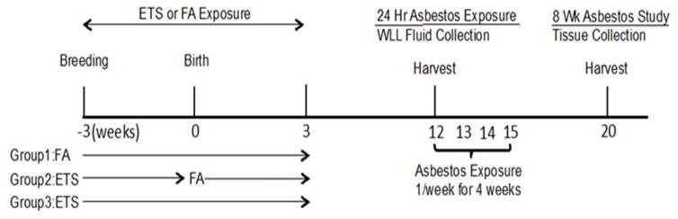
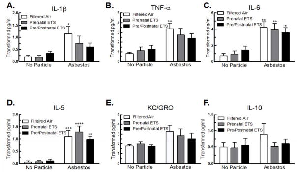
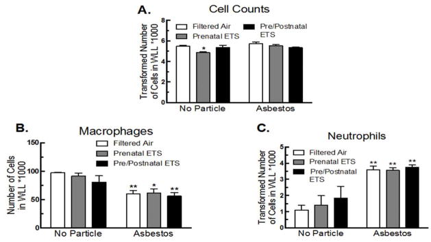
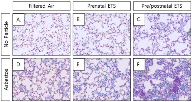
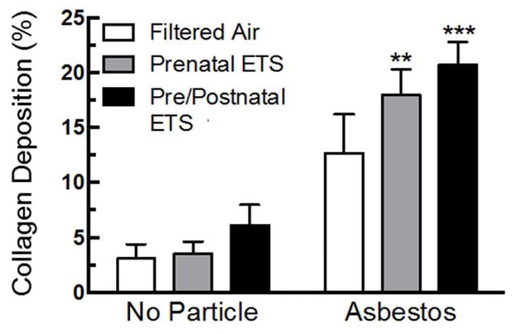
Asbestos in combination with tobacco smoke exposure reportedly leads to more severe physiological consequences than asbestos alone; limited data also show an increased disease risk due to environmental tobacco smoke (ETS) exposure. Environmental influences during gestation and early lung development can result in physiological changes that alter risk for disease development throughout an individual's lifetime. Therefore, maternal lifestyle may impact the ability of offspring to subsequently respond to environmental insults and alter overall disease susceptibility. In this study, we examined the effects of exposure to ETS in utero and during early postnatal development on asbestos-related inflammation and disease in adulthood. ETS exposure in utero appeared to shift inflammation towards a Th2 phenotype, via suppression of Th1 inflammatory cytokine production. This effect was further pronounced in mice exposed to ETS in utero and during early postnatal development. In utero ETS exposure led to increased collagen deposition, a marker of fibrotic disease, when the offspring was later exposed to asbestos, which was further increased with additional ETS exposure during early postnatal development. These data suggest that ETS exposure in utero alters the immune responses and leads to greater disease development after asbestos exposure, which is further exacerbated when exposure to ETS continues during early postnatal development.
Keywords: Asbestos; environmental tobacco smoke; fibrosis; inflammation.
Conflict of interest statement
Declaration of interest No potential conflicts of interest were disclosed.
Figures
Similar articles
-
Prenatal environmental tobacco smoke exposure increases allergic asthma risk with methylation changes in mice.Environ Mol Mutagen. 2017 Jul;58(6):423-433. doi: 10.1002/em.22097. Epub 2017 May 25. Environ Mol Mutagen. 2017. PMID: 28543436 Free PMC article.
-
Effects of environmental tobacco smoke exposure on pulmonary immune response in infant monkeys.J Allergy Clin Immunol. 2008 Aug;122(2):400-6, 406.e1-5. doi: 10.1016/j.jaci.2008.04.011. Epub 2008 May 27. J Allergy Clin Immunol. 2008. PMID: 18502491
-
Prenatal tobacco smoke exposure predisposes offspring mice to exacerbated allergic airway inflammation associated with altered innate effector function.Part Fibre Toxicol. 2017 Aug 22;14(1):30. doi: 10.1186/s12989-017-0212-6. Part Fibre Toxicol. 2017. PMID: 28830530 Free PMC article.
-
Impact of Tobacco Smoke and Nicotine Exposure on Lung Development.Chest. 2016 Feb;149(2):552-561. doi: 10.1378/chest.15-1858. Epub 2016 Jan 12. Chest. 2016. PMID: 26502117 Free PMC article. Review.
-
Detrimental effects of tobacco smoke exposure during development on postnatal lung function and asthma.Birth Defects Res C Embryo Today. 2008 Mar;84(1):54-60. doi: 10.1002/bdrc.20114. Birth Defects Res C Embryo Today. 2008. PMID: 18383132 Review.
Cited by
-
Perinatal exposure to environmental tobacco smoke is associated with changes in DNA methylation that precede the adult onset of lung disease in a mouse model.Inhal Toxicol. 2017 Aug;29(10):435-442. doi: 10.1080/08958378.2017.1392655. Inhal Toxicol. 2017. PMID: 29124997 Free PMC article.
-
Multinucleated giant cell phenotype in response to stimulation.Immunobiology. 2020 May;225(3):151952. doi: 10.1016/j.imbio.2020.151952. Epub 2020 May 5. Immunobiology. 2020. PMID: 32517879 Free PMC article.
-
Methylation-derived Neutrophil-to-Lymphocyte Ratio and Lung Cancer Risk in Heavy Smokers.Cancer Prev Res (Phila). 2018 Nov;11(11):727-734. doi: 10.1158/1940-6207.CAPR-18-0111. Epub 2018 Sep 25. Cancer Prev Res (Phila). 2018. PMID: 30254071 Free PMC article. Clinical Trial.
-
Differential lung inflammation and injury with tobacco smoke exposure in Wistar Kyoto and spontaneously hypertensive rats.Inhal Toxicol. 2020 Jul;32(8):328-341. doi: 10.1080/08958378.2020.1805052. Epub 2020 Aug 11. Inhal Toxicol. 2020. PMID: 32781858 Free PMC article.
-
Assessing health risks from multiple environmental stressors: Moving from G×E to I×E.Mutat Res Rev Mutat Res. 2018 Jan-Mar;775:11-20. doi: 10.1016/j.mrrev.2017.11.003. Epub 2017 Nov 24. Mutat Res Rev Mutat Res. 2018. PMID: 29555026 Free PMC article. Review.
References
-
- ABRAHAMSSON TR, SANDBERG ABELIUS M, FORSBERG A, BJORKSTEN B, JENMALM MC. A Th1/Th2-associated chemokine imbalance during infancy in children developing eczema, wheeze and sensitization. Clin Exp Allergy. 2011;41:1729–39. - PubMed
-
- ARTIS D, SPITS H. The biology of innate lymphoid cells. Nature. 2015;517:293–301. - PubMed
-
- BAUMGARTNER KB, SAMET JM, STIDLEY CA, COLBY TV, WALDRON JA. Cigarette smoking: a risk factor for idiopathic pulmonary fibrosis. Am J Respir Crit Care Med. 1997;155:242–8. - PubMed
Publication types
MeSH terms
Substances
Grants and funding
LinkOut - more resources
Full Text Sources
Other Literature Sources
Medical